• 文献检索
  • 文档翻译
  • 深度研究
  • 学术资讯
  • Suppr Zotero 插件Zotero 插件
  • 邀请有礼
  • 套餐&价格
  • 历史记录
应用&插件
Suppr Zotero 插件Zotero 插件浏览器插件Mac 客户端Windows 客户端微信小程序
定价
高级版会员购买积分包购买API积分包
服务
文献检索文档翻译深度研究API 文档MCP 服务
关于我们
关于 Suppr公司介绍联系我们用户协议隐私条款
关注我们

Suppr 超能文献

核心技术专利:CN118964589B侵权必究
粤ICP备2023148730 号-1Suppr @ 2026

文献检索

告别复杂PubMed语法,用中文像聊天一样搜索,搜遍4000万医学文献。AI智能推荐,让科研检索更轻松。

立即免费搜索

文件翻译

保留排版,准确专业,支持PDF/Word/PPT等文件格式,支持 12+语言互译。

免费翻译文档

深度研究

AI帮你快速写综述,25分钟生成高质量综述,智能提取关键信息,辅助科研写作。

立即免费体验

用于屏障和极性研究的猪单眼视网膜色素上皮细胞培养

Porcine Single-Eye Retinal Pigment Epithelium Cell Culture for Barrier and Polarity Studies.

作者信息

Dörschmann Philipp, von der Weppen Sina, Koyama Emi, Roider Johann, Klettner Alexa

机构信息

Department of Ophthalmology, University Medical Center, Kiel University, Arnold-Heller-Str. 3, Haus B2, 24105 Kiel, Germany.

出版信息

Cells. 2025 Jul 1;14(13):1007. doi: 10.3390/cells14131007.

DOI:10.3390/cells14131007
PMID:40643526
原文链接:https://pmc.ncbi.nlm.nih.gov/articles/PMC12248934/
Abstract

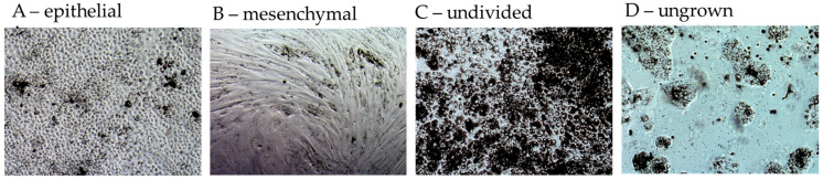
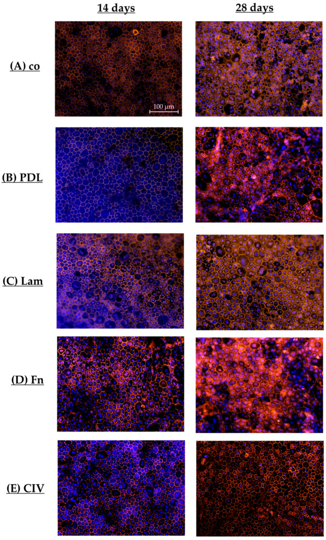
Age-related macular degeneration (AMD) is the main cause of blindness in Western nations. AMD models addressing specific pathological pathways are desired. Through this study, a best-practice protocol for polarized porcine single-eye retinal pigment epithelium (RPE) preparation for AMD-relevant models of RPE barrier and polarity is established. Single-eye porcine primary RPE cells (from one eye for one well) were prepared in 12-well plates including Transwell inserts. Different coatings (laminin (Lam), Poly-ᴅ-Lysine (PDL), fibronectin (Fn) and collagens) and varying serum contents (1%, 5% and 10%) were investigated to determine optimal culture parameters for this model. Success rates of cultures, cell number (trypan-blue exclusion assay), morphology/morphometry (light and fluorescence microscopy), protein secretion/expression (ELISA, Western blot), gene expression (qPCR), transepithelial electric resistance (TEER) and polar location of bestrophin 1 (BEST1) by cryosectioning (IHC-Fr) were assessed. Cells seeded on Lam exhibited the highest level of epithelial cells and confluence properties. Fn resulted in the highest cell number growth. Lam and Fn exhibited the highest culture success rates. TEER values and vascular endothelial growth factor secretion were highest when Lam was used. For the first time, polar (Transwell) porcine single-eye RPE morphometry parameters were determined. RPE on Lam showed bigger cells with a higher variety of cell shapes. CIV displayed the lowest claudin 19 expression. The highest basolateral expression of BEST1 was achieved with Lam coating. The higher the serum, the better the cell number increase and confluence success. A reduction in serum on Lam showed positive results for RPE morphology, while morphometry remained stable. A five percent serum on Lam showed the highest culture success rate and best barrier properties. RPE65 expression was reduced by using 10% serum. Altogether, the most suitable coating of Transwell inserts was Lam, and a reduction in serum to 5% is recommended, as well as a cultivation time of 28 days. A protocol for the use of polar porcine single-eye cultures with validated parameters was established and is provided herein.

摘要

年龄相关性黄斑变性(AMD)是西方国家失明的主要原因。人们期望有针对特定病理途径的AMD模型。通过本研究,建立了一种用于制备与AMD相关的视网膜色素上皮(RPE)屏障和极性模型的偏振猪单眼视网膜色素上皮制备的最佳实践方案。在包括Transwell小室的12孔板中制备单眼猪原代RPE细胞(每孔来自一只眼睛)。研究了不同的包被(层粘连蛋白(Lam)、聚-D-赖氨酸(PDL)、纤连蛋白(Fn)和胶原蛋白)和不同的血清含量(1%、5%和10%),以确定该模型的最佳培养参数。评估培养成功率、细胞数量(台盼蓝排斥试验)、形态/形态测量(光学和荧光显微镜)、蛋白质分泌/表达(ELISA、蛋白质印迹法)、基因表达(qPCR)、跨上皮电阻(TEER)以及通过冷冻切片(免疫组化-Fr)检测的最佳rophin 1(BEST1)的极性定位。接种在Lam上的细胞表现出最高水平的上皮细胞和汇合特性。Fn导致最高的细胞数量增长。Lam和Fn表现出最高的培养成功率。使用Lam时,TEER值和血管内皮生长因子分泌最高。首次确定了极性(Transwell)猪单眼RPE形态测量参数。Lam上的RPE显示细胞更大,细胞形状种类更多样。IV型胶原蛋白显示claudin 19表达最低。使用Lam包被时,BEST1的基底外侧表达最高。血清越高,细胞数量增加和汇合成功率越好。Lam上血清减少对RPE形态显示出积极结果,而形态测量保持稳定。Lam上5%的血清显示出最高的培养成功率和最佳的屏障特性。使用10%血清会降低RPE65表达。总之,Transwell小室最合适的包被是Lam,建议将血清减少到5%,以及培养时间为28天。本文建立并提供了一种使用具有验证参数的极性猪单眼培养物的方案。

https://cdn.ncbi.nlm.nih.gov/pmc/blobs/fd42/12248934/51e33d259a9b/cells-14-01007-g019.jpg
https://cdn.ncbi.nlm.nih.gov/pmc/blobs/fd42/12248934/be100e54a90a/cells-14-01007-g001.jpg
https://cdn.ncbi.nlm.nih.gov/pmc/blobs/fd42/12248934/189f32c4135f/cells-14-01007-g002.jpg
https://cdn.ncbi.nlm.nih.gov/pmc/blobs/fd42/12248934/63ec3b5db102/cells-14-01007-g003.jpg
https://cdn.ncbi.nlm.nih.gov/pmc/blobs/fd42/12248934/bac1263a6f58/cells-14-01007-g004.jpg
https://cdn.ncbi.nlm.nih.gov/pmc/blobs/fd42/12248934/2976125ca3f8/cells-14-01007-g005.jpg
https://cdn.ncbi.nlm.nih.gov/pmc/blobs/fd42/12248934/11f052c477c6/cells-14-01007-g006.jpg
https://cdn.ncbi.nlm.nih.gov/pmc/blobs/fd42/12248934/20d6efcfa2f3/cells-14-01007-g007.jpg
https://cdn.ncbi.nlm.nih.gov/pmc/blobs/fd42/12248934/14dc31d72c69/cells-14-01007-g008.jpg
https://cdn.ncbi.nlm.nih.gov/pmc/blobs/fd42/12248934/56845c59b8bf/cells-14-01007-g009.jpg
https://cdn.ncbi.nlm.nih.gov/pmc/blobs/fd42/12248934/0d055ac0fb53/cells-14-01007-g010.jpg
https://cdn.ncbi.nlm.nih.gov/pmc/blobs/fd42/12248934/bc6294a079db/cells-14-01007-g011.jpg
https://cdn.ncbi.nlm.nih.gov/pmc/blobs/fd42/12248934/a8418974e45b/cells-14-01007-g012.jpg
https://cdn.ncbi.nlm.nih.gov/pmc/blobs/fd42/12248934/dd7bf6d8c0e8/cells-14-01007-g013.jpg
https://cdn.ncbi.nlm.nih.gov/pmc/blobs/fd42/12248934/0604def7b379/cells-14-01007-g014.jpg
https://cdn.ncbi.nlm.nih.gov/pmc/blobs/fd42/12248934/af99f01c9c59/cells-14-01007-g015.jpg
https://cdn.ncbi.nlm.nih.gov/pmc/blobs/fd42/12248934/3cba56ea2bbf/cells-14-01007-g016.jpg
https://cdn.ncbi.nlm.nih.gov/pmc/blobs/fd42/12248934/9ec85d347684/cells-14-01007-g017.jpg
https://cdn.ncbi.nlm.nih.gov/pmc/blobs/fd42/12248934/838e042db7fd/cells-14-01007-g018.jpg
https://cdn.ncbi.nlm.nih.gov/pmc/blobs/fd42/12248934/51e33d259a9b/cells-14-01007-g019.jpg

相似文献

1
Porcine Single-Eye Retinal Pigment Epithelium Cell Culture for Barrier and Polarity Studies.用于屏障和极性研究的猪单眼视网膜色素上皮细胞培养
Cells. 2025 Jul 1;14(13):1007. doi: 10.3390/cells14131007.
2
Adefovir dipivoxil and pegylated interferon alfa-2a for the treatment of chronic hepatitis B: a systematic review and economic evaluation.阿德福韦酯与聚乙二醇化干扰素α-2a治疗慢性乙型肝炎:系统评价与经济学评估
Health Technol Assess. 2006 Aug;10(28):iii-iv, xi-xiv, 1-183. doi: 10.3310/hta10280.
3
Artificial intelligence for diagnosing exudative age-related macular degeneration.人工智能在渗出性年龄相关性黄斑变性诊断中的应用。
Cochrane Database Syst Rev. 2024 Oct 17;10(10):CD015522. doi: 10.1002/14651858.CD015522.pub2.
4
Influence of carrier materials and coatings on retinal pigment epithelium cultivation and functions.载体材料和涂层对视网膜色素上皮细胞培养和功能的影响。
Exp Eye Res. 2022 Jun;219:109063. doi: 10.1016/j.exer.2022.109063. Epub 2022 Apr 3.
5
Blue-light filtering intraocular lenses (IOLs) for protecting macular health.用于保护黄斑健康的蓝光滤过型人工晶状体
Cochrane Database Syst Rev. 2018 May 22;5(5):CD011977. doi: 10.1002/14651858.CD011977.pub2.
6
Lateral flow urine lipoarabinomannan assay for detecting active tuberculosis in HIV-positive adults.用于检测HIV阳性成年人活动性结核病的侧向流动尿液脂阿拉伯甘露聚糖检测法
Cochrane Database Syst Rev. 2016 May 10;2016(5):CD011420. doi: 10.1002/14651858.CD011420.pub2.
7
Anti-vascular endothelial growth factor biosimilars for neovascular age-related macular degeneration.抗血管内皮生长因子生物类似药治疗新生血管性年龄相关性黄斑变性。
Cochrane Database Syst Rev. 2024 Jun 3;6(6):CD015804. doi: 10.1002/14651858.CD015804.pub2.
8
Interventions for central serous chorioretinopathy: a network meta-analysis.中心性浆液性脉络膜视网膜病变的干预措施:一项网状Meta分析
Cochrane Database Syst Rev. 2025 Jun 16;6(6):CD011841. doi: 10.1002/14651858.CD011841.pub3.
9
Aflibercept for neovascular age-related macular degeneration.阿柏西普用于治疗新生血管性年龄相关性黄斑变性。
Cochrane Database Syst Rev. 2016 Feb 8;2(2):CD011346. doi: 10.1002/14651858.CD011346.pub2.
10
The influence of femtosecond laser intrastromal lenticules on the characteristics and maturity in tissue-engineered stem cell-derived retinal pigment epithelium sheets.飞秒激光基质内透镜对组织工程干细胞源性视网膜色素上皮片的特性及成熟度的影响
Stem Cell Res Ther. 2025 Jun 20;16(1):316. doi: 10.1186/s13287-025-04463-7.

本文引用的文献

1
Recent Achievements in the Heterogeneity of Mammalian and Human Retinal Pigment Epithelium: In Search of a Stem Cell.哺乳动物和人类视网膜色素上皮细胞异质性的最新研究进展:寻找干细胞。
Cells. 2024 Feb 4;13(3):281. doi: 10.3390/cells13030281.
2
Subconfluent ARPE-19 Cells Display Mesenchymal Cell-State Characteristics and Behave like Fibroblasts, Rather Than Epithelial Cells, in Experimental HCMV Infection Studies.亚汇合 ARPE-19 细胞在实验性 HCMV 感染研究中表现出间充质细胞状态特征,并表现为成纤维细胞,而不是上皮细胞。
Viruses. 2023 Dec 28;16(1):49. doi: 10.3390/v16010049.
3
Identifying biomarkers of heterogeneity and transplantation efficacy in retinal pigment epithelial cells.
鉴定视网膜色素上皮细胞异质性和移植疗效的生物标志物。
J Exp Med. 2023 Dec 4;220(12). doi: 10.1084/jem.20230913. Epub 2023 Sep 20.
4
Transcriptional comparison of adult human primary Retinal Pigment Epithelium, human pluripotent stem cell-derived Retinal Pigment Epithelium, and ARPE19 cells.成人人类原代视网膜色素上皮细胞、人多能干细胞衍生的视网膜色素上皮细胞和ARPE19细胞的转录比较
Front Cell Dev Biol. 2022 Aug 26;10:910040. doi: 10.3389/fcell.2022.910040. eCollection 2022.
5
Differentiation and Maturation Effect of All-Trans Retinoic Acid on Cultured Fetal RPE and Stem Cell-Derived RPE Cells for Cell-Based Therapy.全反式视黄酸对培养的胎儿 RPE 和干细胞衍生的 RPE 细胞用于细胞治疗的分化和成熟作用。
Curr Eye Res. 2022 Sep;47(9):1300-1311. doi: 10.1080/02713683.2022.2079144. Epub 2022 Jul 27.
6
Multinucleated Retinal Pigment Epithelial Cells Adapt to Vision and Exhibit Increased DNA Damage Response.多核视网膜色素上皮细胞适应视觉并表现出增强的 DNA 损伤反应。
Cells. 2022 May 5;11(9):1552. doi: 10.3390/cells11091552.
7
Influence of carrier materials and coatings on retinal pigment epithelium cultivation and functions.载体材料和涂层对视网膜色素上皮细胞培养和功能的影响。
Exp Eye Res. 2022 Jun;219:109063. doi: 10.1016/j.exer.2022.109063. Epub 2022 Apr 3.
8
Culture surface protein coatings affect the barrier properties and calcium signalling of hESC-RPE.培养表面蛋白涂层会影响 hESC-RPE 的屏障特性和钙信号转导。
Sci Rep. 2021 Jan 13;11(1):933. doi: 10.1038/s41598-020-79638-8.
9
Basolateral activation with TLR agonists induces polarized cytokine release and reduces barrier function in RPE in vitro.TLR 激动剂诱导基底外侧激活,导致体外 RPE 极化细胞因子释放和屏障功能降低。
Graefes Arch Clin Exp Ophthalmol. 2021 Feb;259(2):413-424. doi: 10.1007/s00417-020-04930-2. Epub 2020 Sep 19.
10
Retina in a dish: Cell cultures, retinal explants and animal models for common diseases of the retina.离体视网膜:用于研究常见视网膜疾病的细胞培养、视网膜组织块和动物模型。
Prog Retin Eye Res. 2021 Mar;81:100880. doi: 10.1016/j.preteyeres.2020.100880. Epub 2020 Jul 25.