• 文献检索
  • 文档翻译
  • 深度研究
  • 学术资讯
  • Suppr Zotero 插件Zotero 插件
  • 邀请有礼
  • 套餐&价格
  • 历史记录
应用&插件
Suppr Zotero 插件Zotero 插件浏览器插件Mac 客户端Windows 客户端微信小程序
定价
高级版会员购买积分包购买API积分包
服务
文献检索文档翻译深度研究API 文档MCP 服务
关于我们
关于 Suppr公司介绍联系我们用户协议隐私条款
关注我们

Suppr 超能文献

核心技术专利:CN118964589B侵权必究
粤ICP备2023148730 号-1Suppr @ 2026

文献检索

告别复杂PubMed语法,用中文像聊天一样搜索,搜遍4000万医学文献。AI智能推荐,让科研检索更轻松。

立即免费搜索

文件翻译

保留排版,准确专业,支持PDF/Word/PPT等文件格式,支持 12+语言互译。

免费翻译文档

深度研究

AI帮你快速写综述,25分钟生成高质量综述,智能提取关键信息,辅助科研写作。

立即免费体验

蓝光和绿光眼底自发荧光在年龄相关性黄斑变性评估中对光学相干断层扫描的补充作用

Blue Light and Green Light Fundus Autofluorescence, Complementary to Optical Coherence Tomography, in Age-Related Macular Degeneration Evaluation.

作者信息

Ranetti Antonia-Elena, Stanca Horia Tudor, Munteanu Mihnea, Bievel Radulescu Raluca, Stanca Simona

机构信息

Doctoral School, "Carol Davila" University of Medicine and Pharmacy, Strada Dionisie Lupu No. 37, 020021 București, Romania.

Clinical Department of Ophthalmology, "Carol Davila" University of Medicine and Pharmacy, 020021 Bucharest, Romania.

出版信息

Diagnostics (Basel). 2025 Jul 2;15(13):1688. doi: 10.3390/diagnostics15131688.

DOI:10.3390/diagnostics15131688
PMID:40647687
原文链接:https://pmc.ncbi.nlm.nih.gov/articles/PMC12248692/
Abstract

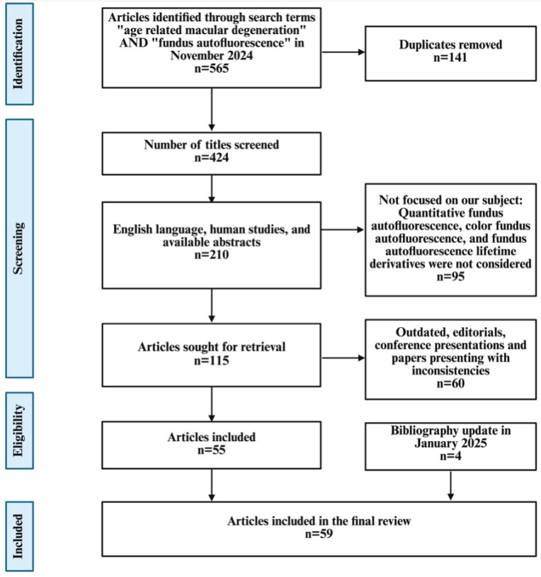
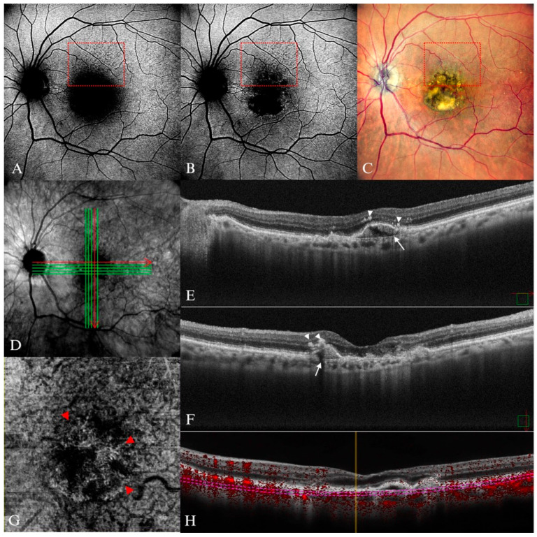
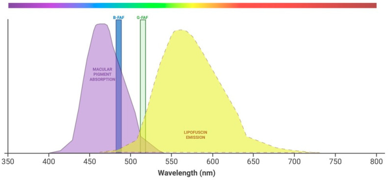
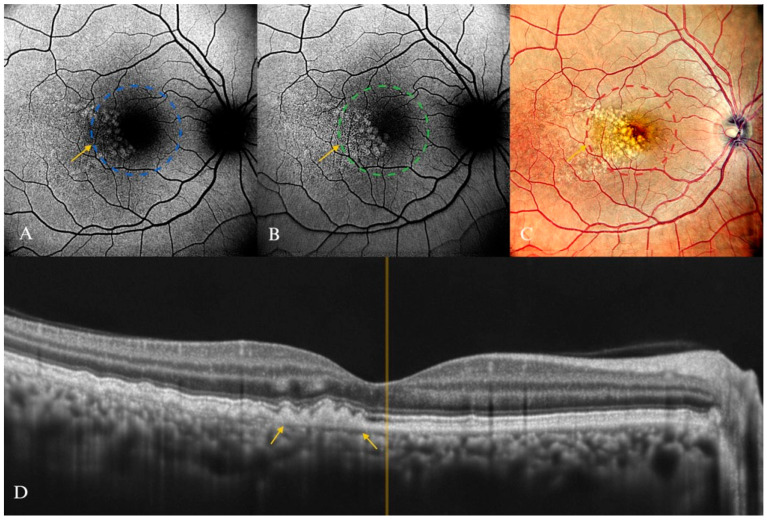
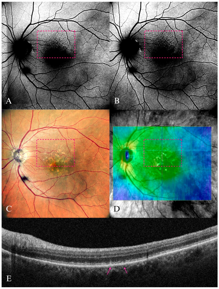
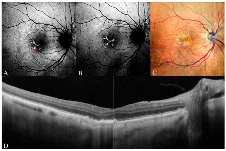
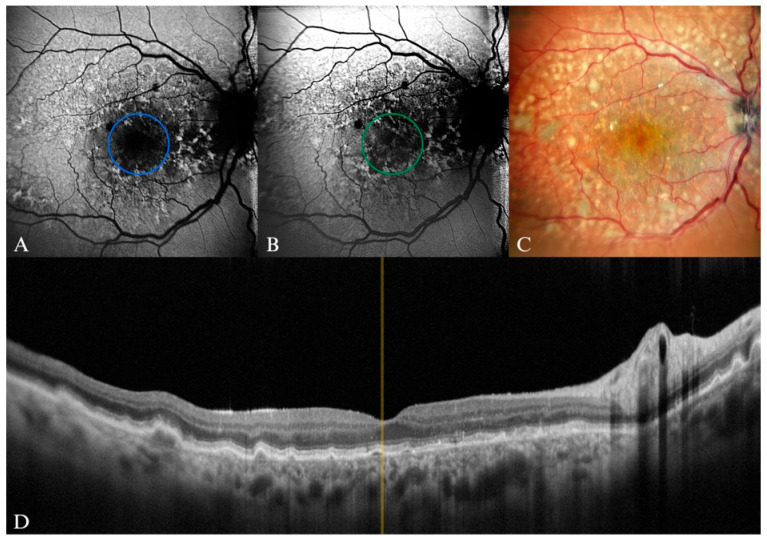
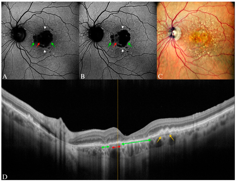
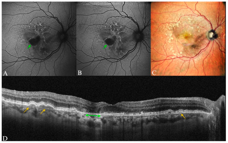
Age-related macular degeneration (AMD) is one of the leading causes of permanent vision loss in the elderly, particularly in higher-income countries. Fundus autofluorescence (FAF) imaging is a widely used, non-invasive technique that complements structural imaging in the assessment of retinal pigment epithelium (RPE) integrity. While optical coherence tomography (OCT) remains the gold standard for retinal imaging due to its high-resolution cross-sectional visualization, FAF offers unique metabolic insights. Among the FAF modalities, blue light FAF (B-FAF) is more commonly employed, whereas green light FAF (G-FAF) provides subtly different image characteristics, particularly improved visualization and contrast in the central macula. Despite identical acquisition times and nearly indistinguishable workflows, G-FAF is notably underutilized in clinical practice. This narrative review critically compares green and blue FAF in terms of their diagnostic utility relative to OCT, with a focus on lesion detectability, macular pigment interference, and clinical decision-making in retinal disorders. A comprehensive literature search was performed using the PubMed database for studies published prior to February 2025. The search utilized the keywords fundus autofluorescence and age-related macular degeneration. The primary focus was on short-wavelength FAF and its clinical utility in AMD, considering three aspects: diagnosis, follow-up, and prognosis. The OCT findings served as the reference standard for anatomical correlation and diagnostic accuracy. Both FAF modalities correlated well with OCT in detecting RPE abnormalities. G-FAF demonstrated improved visibility of central lesions due to reduced masking by macular pigment and enhanced contrast in the macula. However, clinical preference remained skewed toward B-FAF, driven more by tradition and device default settings than by evidence-based superiority. G-FAF's diagnostic potential remains underrecognized despite its comparable practicality and subtle imaging advantages specifically for AMD patients. AMD stages were accurately characterized, and relevant images were used to highlight the significance of G-FAF and B-FAF in the examination of AMD patients. While OCT remains the gold standard, FAF provides complementary information that can guide management strategy. Since G-FAF is functionally equivalent in acquisition, it offers slight advantages. Broader awareness and more frequent integration of G-FAF that could optimize multimodal imaging strategies, particularly in the intermediate stage, should be developed.

摘要

年龄相关性黄斑变性(AMD)是老年人永久性视力丧失的主要原因之一,在高收入国家尤其如此。眼底自发荧光(FAF)成像是一种广泛使用的非侵入性技术,在评估视网膜色素上皮(RPE)完整性方面可补充结构成像。虽然光学相干断层扫描(OCT)因其高分辨率的横断面可视化仍然是视网膜成像的金标准,但FAF提供了独特的代谢见解。在FAF模式中,蓝光FAF(B-FAF)更常用,而绿光FAF(G-FAF)提供略有不同的图像特征,特别是在黄斑中心区改善了可视化和对比度。尽管采集时间相同且工作流程几乎难以区分,但G-FAF在临床实践中的应用明显不足。本叙述性综述严格比较了绿色和蓝色FAF相对于OCT的诊断效用,重点关注病变可检测性、黄斑色素干扰以及视网膜疾病的临床决策。使用PubMed数据库对2025年2月之前发表的研究进行了全面的文献检索。检索使用了关键词“眼底自发荧光”和“年龄相关性黄斑变性”。主要关注短波长FAF及其在AMD中的临床效用,考虑三个方面:诊断、随访和预后。OCT检查结果作为解剖相关性和诊断准确性的参考标准。两种FAF模式在检测RPE异常方面与OCT相关性良好。由于黄斑色素的掩盖减少以及黄斑区对比度增强,G-FAF显示出中央病变的可见性提高。然而,临床偏好仍然倾向于B-FAF,更多是受传统和设备默认设置的驱动,而非基于证据的优越性。尽管G-FAF具有相当的实用性和对AMD患者特别的细微成像优势,但其诊断潜力仍未得到充分认识。准确描述了AMD的阶段,并使用相关图像突出了G-FAF和B-FAF在AMD患者检查中的重要性。虽然OCT仍然是金标准,但FAF提供了可指导管理策略的补充信息。由于G-FAF在采集功能上等效,它具有一些轻微优势。应提高对G-FAF的更广泛认识并更频繁地将其纳入,以优化多模态成像策略,特别是在中期阶段。

https://cdn.ncbi.nlm.nih.gov/pmc/blobs/db31/12248692/0bc31ee5396f/diagnostics-15-01688-g014.jpg
https://cdn.ncbi.nlm.nih.gov/pmc/blobs/db31/12248692/dcc2414e2d35/diagnostics-15-01688-g001.jpg
https://cdn.ncbi.nlm.nih.gov/pmc/blobs/db31/12248692/d897676483b2/diagnostics-15-01688-g002.jpg
https://cdn.ncbi.nlm.nih.gov/pmc/blobs/db31/12248692/f785277ec8fb/diagnostics-15-01688-g003.jpg
https://cdn.ncbi.nlm.nih.gov/pmc/blobs/db31/12248692/ed845d9f25bc/diagnostics-15-01688-g004.jpg
https://cdn.ncbi.nlm.nih.gov/pmc/blobs/db31/12248692/f7d8a9f56423/diagnostics-15-01688-g005.jpg
https://cdn.ncbi.nlm.nih.gov/pmc/blobs/db31/12248692/0a2e6687e572/diagnostics-15-01688-g006.jpg
https://cdn.ncbi.nlm.nih.gov/pmc/blobs/db31/12248692/86221cfda7a3/diagnostics-15-01688-g007.jpg
https://cdn.ncbi.nlm.nih.gov/pmc/blobs/db31/12248692/caf1ce9ef7e9/diagnostics-15-01688-g008.jpg
https://cdn.ncbi.nlm.nih.gov/pmc/blobs/db31/12248692/6660e4c455a7/diagnostics-15-01688-g009.jpg
https://cdn.ncbi.nlm.nih.gov/pmc/blobs/db31/12248692/a3c3272373c3/diagnostics-15-01688-g010.jpg
https://cdn.ncbi.nlm.nih.gov/pmc/blobs/db31/12248692/d02de3fb3afb/diagnostics-15-01688-g011.jpg
https://cdn.ncbi.nlm.nih.gov/pmc/blobs/db31/12248692/b867144a9eb6/diagnostics-15-01688-g012.jpg
https://cdn.ncbi.nlm.nih.gov/pmc/blobs/db31/12248692/ab396713cec0/diagnostics-15-01688-g013.jpg
https://cdn.ncbi.nlm.nih.gov/pmc/blobs/db31/12248692/0bc31ee5396f/diagnostics-15-01688-g014.jpg

相似文献

1
Blue Light and Green Light Fundus Autofluorescence, Complementary to Optical Coherence Tomography, in Age-Related Macular Degeneration Evaluation.蓝光和绿光眼底自发荧光在年龄相关性黄斑变性评估中对光学相干断层扫描的补充作用
Diagnostics (Basel). 2025 Jul 2;15(13):1688. doi: 10.3390/diagnostics15131688.
2
Optical coherence tomography (OCT) for detection of macular oedema in patients with diabetic retinopathy.光学相干断层扫描(OCT)用于检测糖尿病视网膜病变患者的黄斑水肿。
Cochrane Database Syst Rev. 2015 Jan 7;1(1):CD008081. doi: 10.1002/14651858.CD008081.pub3.
3
Optical coherence tomography (OCT) for detection of macular oedema in patients with diabetic retinopathy.光学相干断层扫描(OCT)用于检测糖尿病视网膜病变患者的黄斑水肿。
Cochrane Database Syst Rev. 2011 Jul 6(7):CD008081. doi: 10.1002/14651858.CD008081.pub2.
4
Artificial intelligence for diagnosing exudative age-related macular degeneration.人工智能在渗出性年龄相关性黄斑变性诊断中的应用。
Cochrane Database Syst Rev. 2024 Oct 17;10(10):CD015522. doi: 10.1002/14651858.CD015522.pub2.
5
Accuracy of fundus autofluorescence imaging for the diagnosis and monitoring of retinal conditions: a systematic review.眼底自发荧光成像在视网膜疾病诊断和监测中的准确性:一项系统评价
Health Technol Assess. 2016 Apr;20(31):1-108. doi: 10.3310/hta20310.
6
Blue-light filtering intraocular lenses (IOLs) for protecting macular health.用于保护黄斑健康的蓝光滤过型人工晶状体
Cochrane Database Syst Rev. 2018 May 22;5(5):CD011977. doi: 10.1002/14651858.CD011977.pub2.
7
Aflibercept for neovascular age-related macular degeneration.阿柏西普用于治疗新生血管性年龄相关性黄斑变性。
Cochrane Database Syst Rev. 2016 Feb 8;2(2):CD011346. doi: 10.1002/14651858.CD011346.pub2.
8
Management of urinary stones by experts in stone disease (ESD 2025).结石病专家对尿路结石的管理(2025年结石病专家共识)
Arch Ital Urol Androl. 2025 Jun 30;97(2):14085. doi: 10.4081/aiua.2025.14085.
9
Artificial intelligence for detecting keratoconus.人工智能在圆锥角膜检测中的应用。
Cochrane Database Syst Rev. 2023 Nov 15;11(11):CD014911. doi: 10.1002/14651858.CD014911.pub2.
10
123I-MIBG scintigraphy and 18F-FDG-PET imaging for diagnosing neuroblastoma.用于诊断神经母细胞瘤的123I-间碘苄胍闪烁扫描术和18F-氟代脱氧葡萄糖正电子发射断层显像
Cochrane Database Syst Rev. 2015 Sep 29;2015(9):CD009263. doi: 10.1002/14651858.CD009263.pub2.

本文引用的文献

1
Age-Related Macular Degeneration Preferred Practice Pattern®.年龄相关性黄斑变性首选诊疗模式®
Ophthalmology. 2025 Apr;132(4):P1-P74. doi: 10.1016/j.ophtha.2024.12.018. Epub 2025 Feb 7.
2
Fundus Autofluorescence Variation in Geographic Atrophy of Age-Related Macular Degeneration: A Clinicopathologic Correlation.年龄相关性黄斑变性地图样萎缩中的眼底自发荧光变化:临床病理相关性研究
Invest Ophthalmol Vis Sci. 2025 Jan 2;66(1):49. doi: 10.1167/iovs.66.1.49.
3
Photoreceptor-RPE loss ratio and fundus autofluorescence patterns as predictive factors for lesion progression in geographic atrophy.
光感受器-视网膜色素上皮细胞损失率和眼底自发荧光模式作为地图样萎缩病变进展的预测因素。
Acta Ophthalmol. 2025 Jun;103(4):e204-e212. doi: 10.1111/aos.17431. Epub 2024 Dec 18.
4
Association of Hyperautofluorescence Signals with Geographic Atrophy Progression in the METformin for the MINimization of Geographic Atrophy Progression Trial.二甲双胍最小化地图样萎缩进展试验中高自发荧光信号与地图样萎缩进展的关联
Ophthalmol Sci. 2024 Sep 12;5(1):100620. doi: 10.1016/j.xops.2024.100620. eCollection 2025 Jan-Feb.
5
Comparison of Geographic Atrophy Measurements Between Blue-Light Heidelberg Standard Field and Green-Light Optos Ultrawide Field Autofluorescence.蓝光源海德堡标准视野和绿光源 Optos 超广角荧光素自发成像测量地理萎缩的比较。
Transl Vis Sci Technol. 2024 Nov 4;13(11):1. doi: 10.1167/tvst.13.11.1.
6
Correlation Between Blue Fundus Autofluorescence and SD-OCT Measurements of Geographic Atrophy in Dry Age-Related Macular Degeneration.干性年龄相关性黄斑变性中蓝色眼底自发荧光与 SD-OCT 测量的地理萎缩的相关性。
Am J Ophthalmol. 2024 Oct;266:92-101. doi: 10.1016/j.ajo.2024.04.031. Epub 2024 May 7.
7
Comparison of Blue-Light Autofluorescence and Ultrawidefield Green-Light Autofluorescence for Assessing Geographic Atrophy.比较蓝光自发荧光和超广角绿光自发荧光在评估地图状萎缩中的应用。
Ophthalmol Retina. 2024 Oct;8(10):987-993. doi: 10.1016/j.oret.2024.04.017. Epub 2024 Apr 25.
8
Age-Related Macular Degeneration: A Review.年龄相关性黄斑变性:综述。
JAMA. 2024 Jan 9;331(2):147-157. doi: 10.1001/jama.2023.26074.
9
Fundus Autofluorescence Patterns in Subretinal Hemorrhages Associated with Neovascular Age-Related Macular Degeneration.视网膜下出血与新生血管性年龄相关性黄斑变性相关的眼底自发荧光模式。
Ophthalmologica. 2024;247(1):58-64. doi: 10.1159/000535865. Epub 2023 Dec 19.
10
Pegcetacoplan for the treatment of geographic atrophy secondary to age-related macular degeneration (OAKS and DERBY): two multicentre, randomised, double-masked, sham-controlled, phase 3 trials.培格司他单抗治疗与年龄相关的黄斑变性(OAKS 和 DERBY)相关的地图状萎缩:两项多中心、随机、双盲、假对照、3 期临床试验。
Lancet. 2023 Oct 21;402(10411):1434-1448. doi: 10.1016/S0140-6736(23)01520-9.