Jlailati Alaa, Al Sbenaty Ghazal, Boali Osama, Younes Deema, Alhayek Bakr, Mozi Baraah, Al-Bitar Ahmad, Bakkour Moudar
Faculty of Medicine, Damascus University, Damascus, Syrian Arab Republic.
Department of Internal Medicine, University of South Florida, Tampa, USA.
J Med Case Rep. 2025 Jul 12;19(1):339. doi: 10.1186/s13256-025-05368-y.
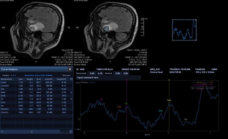
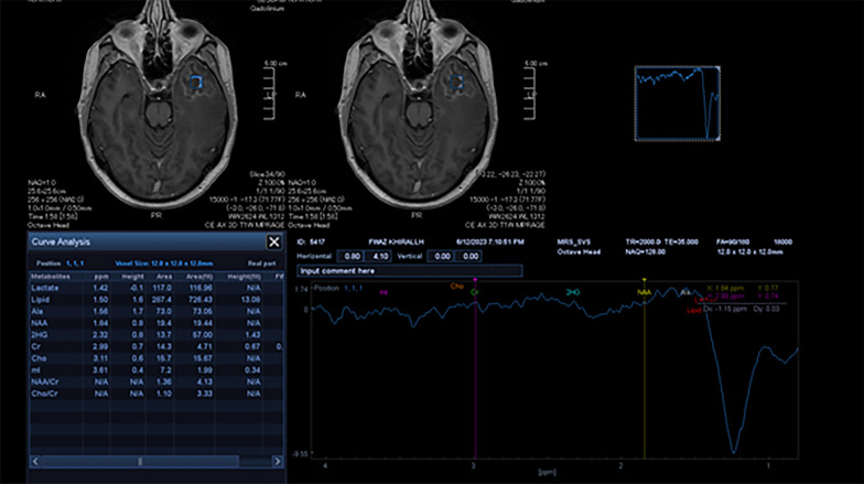
Nasopharyngeal carcinoma is considered rare worldwide. The treatment of nasopharyngeal carcinoma primarily relies on radiotherapy, as the tumor cells in NPC exhibit radiosensitivity. However, excessive dosage can result in a delayed reaction that affects the healthy surrounding tissues, including the central nervous system, causing brain radionecrosis, a rare yet severe condition, which can develop approximately 6-12 months after radiation as a significant complication. The majority of studies on brain radionecrosis have been conducted in China, where nasopharyngeal carcinoma is most prevalent. However, to the best of our knowledge, this is the first reported case of brain radionecrosis following radiotherapy for nasopharyngeal carcinoma in our region, which was diagnosed using magnetic resonance spectroscopy.
This case report describes a 49-year-old Arab male who presented with memory loss, expressive aphasia, and delirium 5 months after undergoing radiotherapy with a total radiation dose of 66 Gray for nasopharyngeal carcinoma (NPC). Magnetic resonance imaging with magnetic resonance spectroscopy revealed the presence of focal lesions in the left temporal lobe with accompanying brain edema indicative of radionecrosis.
It is imperative to consider the possibility of brain radionecrosis in patients who have previously received radiation therapy for head and neck cancers, particularly nasopharyngeal carcinoma. Early detection of brain radionecrosis is essential, and diagnostic imaging should be performed regularly during follow-up using magnetic resonance imaging and magnetic resonance spectroscopy. The primary objective of treatment is to alleviate symptoms through medical and/or surgical interventions.
鼻咽癌在全球范围内被认为较为罕见。鼻咽癌的治疗主要依赖放疗,因为鼻咽癌的肿瘤细胞具有放射敏感性。然而,过量的剂量会导致延迟反应,影响周围健康组织,包括中枢神经系统,引发脑放射性坏死,这是一种罕见但严重的病症,可在放疗后约6至12个月作为一种重大并发症出现。大多数关于脑放射性坏死的研究是在中国进行的,那里鼻咽癌最为普遍。然而,据我们所知,这是我们地区首例报告的鼻咽癌放疗后脑放射性坏死病例,该病例通过磁共振波谱进行了诊断。
本病例报告描述了一名49岁的阿拉伯男性,他在接受了总辐射剂量为66戈瑞的鼻咽癌放疗5个月后,出现了记忆力减退、表达性失语和谵妄症状。磁共振成像结合磁共振波谱显示左颞叶存在局灶性病变,并伴有提示放射性坏死的脑水肿。
对于先前接受过头颈部癌症放疗,尤其是鼻咽癌放疗的患者,必须考虑脑放射性坏死的可能性。早期发现脑放射性坏死至关重要,在随访期间应定期使用磁共振成像和磁共振波谱进行诊断性成像。治疗的主要目标是通过药物和/或手术干预缓解症状。