Golinska Monika, Rycerz Aleksander, Sobczak Matylda, Chrzanowski Jedrzej, Stawiski Konrad, Fendler Wojciech
Department of Biostatistics and Translational Medicine, Medical University of Lodz, Lodz, Poland.
Cancer Research UK Cambridge Institute (CRUK CI), University of Cambridge, Cambridge, United Kingdom.
Front Immunol. 2025 Jul 8;16:1619434. doi: 10.3389/fimmu.2025.1619434. eCollection 2025.
Molecular events that drive endometriosis (EM) and cause accompanying immune deregulation remain elusive. Our purpose was to identify key pathways involved in lesion formation across diverse populations and to detect transcriptomic changes in eutopic endometrium that accompany EM.
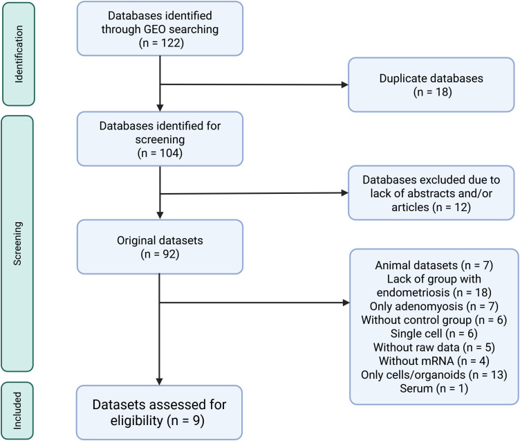
We searched Gene Expression Omnibus and ArrayExpress and performed differential gene expression analysis and a network meta-analysis on nine qualifying datasets. Those contained transcriptomic data on 114 ectopic endometrium samples (EL), 138 eutopic endometrium samples from women with endometriosis (EEM), and 79 eutopic endometrium samples from women without endometriosis (EH). Gene ontology and enrichment analysis were performed in DAVID, Metascape, and Cytoscape, and drug repurposing was done in CMap.
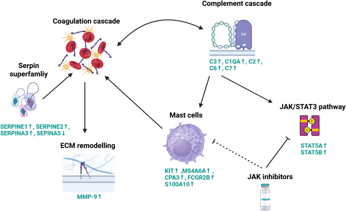
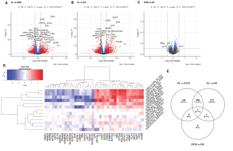
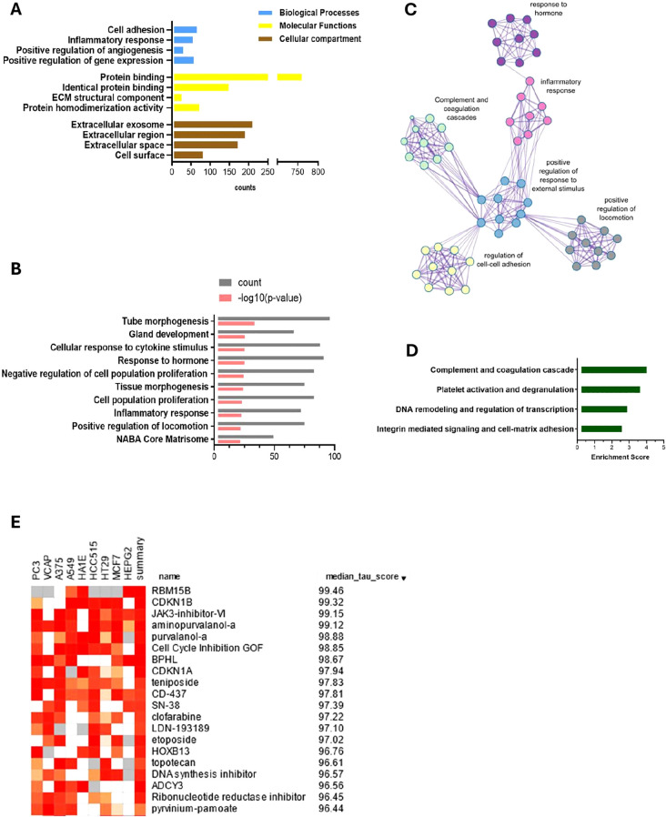
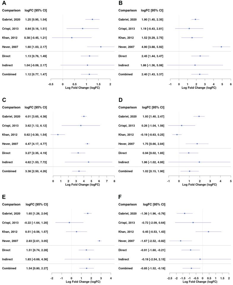
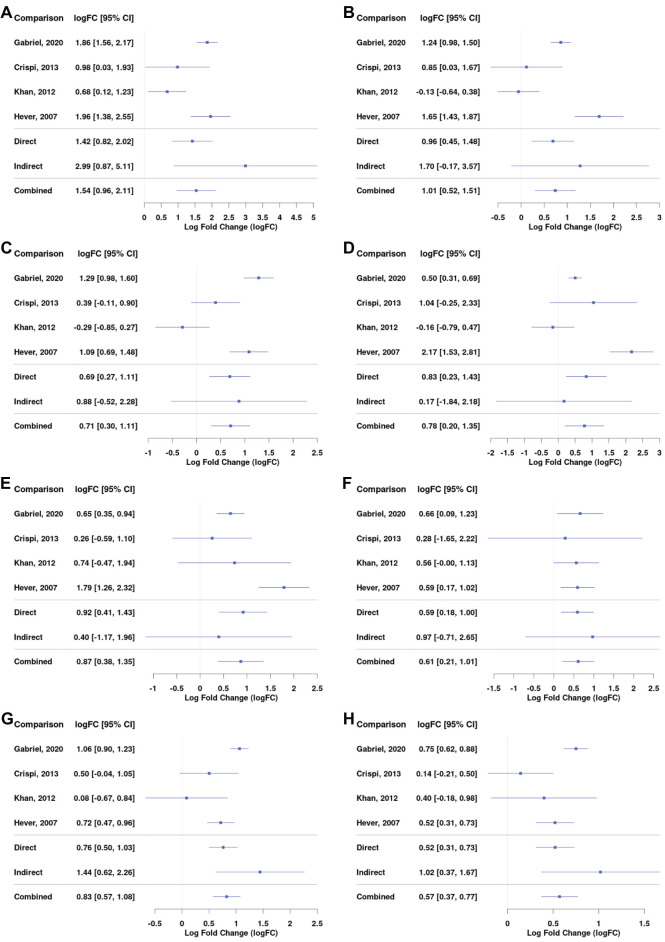
EEM compared to EH upregulated and downregulated , , and genes (|logFC| > 0.5, < 0.05). EL showed increased expression of complement and serpin genes (EL vs. EEM: , logFC = 3.38, < 0.0001; , logFC = 2.40, < 0.0001; , logFC = 1.02, < 0.05; , logFC = 1.54, < 0.001) and mast cell markers (EL vs. EEM: , logFC = 1.54, < 0.0001; , logFC = 0.74, < 0.001). Functional enrichment analysis highlighted complement and coagulation, inflammation, angiogenesis, and extracellular matrix remodeling as drivers of endometriosis. Pharmacogenomic analysis indicated Janus kinase (JAK), cyclin-dependent kinase (CDK), and topoisomerase inhibitors as therapy targets.
Our results suggest an interplay between complement and coagulation, mast cells, extracellular matrix remodeling, and the JAK/STAT3 pathway in endometriosis. We underscore the significance of complement C3 and propose JAK inhibitors as therapy candidates. Detected expression differences between EEM and EH are important for the development of diagnosis via endometrial biopsy.
驱动子宫内膜异位症(EM)并导致伴随免疫失调的分子事件仍不清楚。我们的目的是确定不同人群中参与病变形成的关键途径,并检测伴随EM的在位子宫内膜的转录组变化。
我们检索了基因表达综合数据库(Gene Expression Omnibus)和ArrayExpress,并对九个合格数据集进行了差异基因表达分析和网络荟萃分析。这些数据集包含114个异位子宫内膜样本(EL)、138个来自子宫内膜异位症患者的在位子宫内膜样本(EEM)和79个来自无子宫内膜异位症女性的在位子宫内膜样本(EH)的转录组数据。在DAVID、Metascape和Cytoscape中进行基因本体和富集分析,并在CMap中进行药物再利用分析。
与EH相比,EEM上调和下调了、、和基因(|logFC|>0.5,<0.05)。EL显示补体和丝氨酸蛋白酶抑制剂基因表达增加(EL与EEM相比:,logFC = 3.38,<0.0001;,logFC = 2.40,<0.0001;,logFC = 1.02,<0.05;,logFC = 1.54,<0.001)以及肥大细胞标志物(EL与EEM相比:,logFC = 1.54,<0.0001;,logFC = 0.74,<0.001)。功能富集分析突出了补体和凝血、炎症、血管生成以及细胞外基质重塑是子宫内膜异位症的驱动因素。药物基因组学分析表明 Janus激酶(JAK)、细胞周期蛋白依赖性激酶(CDK)和拓扑异构酶抑制剂是治疗靶点。
我们的结果表明补体与凝血、肥大细胞、细胞外基质重塑以及JAK/STAT3途径在子宫内膜异位症中相互作用。我们强调补体C3的重要性,并提出JAK抑制剂作为治疗候选药物。检测到的EEM和EH之间的表达差异对于通过子宫内膜活检进行诊断的发展很重要。