• 文献检索
  • 文档翻译
  • 深度研究
  • 学术资讯
  • Suppr Zotero 插件Zotero 插件
  • 邀请有礼
  • 套餐&价格
  • 历史记录
应用&插件
Suppr Zotero 插件Zotero 插件浏览器插件Mac 客户端Windows 客户端微信小程序
定价
高级版会员购买积分包购买API积分包
服务
文献检索文档翻译深度研究API 文档MCP 服务
关于我们
关于 Suppr公司介绍联系我们用户协议隐私条款
关注我们

Suppr 超能文献

核心技术专利:CN118964589B侵权必究
粤ICP备2023148730 号-1Suppr @ 2026

文献检索

告别复杂PubMed语法,用中文像聊天一样搜索,搜遍4000万医学文献。AI智能推荐,让科研检索更轻松。

立即免费搜索

文件翻译

保留排版,准确专业,支持PDF/Word/PPT等文件格式,支持 12+语言互译。

免费翻译文档

深度研究

AI帮你快速写综述,25分钟生成高质量综述,智能提取关键信息,辅助科研写作。

立即免费体验

神经节细胞层厚度作为肌萎缩侧索硬化症功能预后的生物标志物:一项光学相干断层扫描研究。

Ganglion Cell Layer Thickness as a Biomarker for Amyotrophic Lateral Sclerosis Functional Outcome: An OCT study.

作者信息

Singh Divya, Singhal Somya, Kanaujiya Vikas, Ranjan Ankita, Mani Vinita Elizabeth, Paliwal Vimal Kumar, Jain Vaibhav, Aishwarya Ankita, Agarwal Rachna

机构信息

Department of Ophthalmology, Sanjay Gandhi Post Graduate Institute of Medical Sciences, Lucknow, India.

Department of Neurology, Sanjay Gandhi Post Graduate Institute of Medical Sciences, Lucknow, India.

出版信息

Rom J Ophthalmol. 2025 Apr-Jun;69(2):200-207. doi: 10.22336/rjo.2025.32.

DOI:10.22336/rjo.2025.32
PMID:40698100
原文链接:https://pmc.ncbi.nlm.nih.gov/articles/PMC12277982/
Abstract

AIM

This study aims to evaluate various optical coherence tomography (OCT) parameters in patients diagnosed with amyotrophic lateral sclerosis (ALS).

METHODS

Assessment of BCVA was done using Snellen charts, and subjective refraction was done to achieve a BCVA for distance and near. Measurement of intraocular pressure (IOP) was done with Goldman applanation tonometry. Stereoscopic fundus examination was performed using a 90D lens to assess the status of the optic nerve and retina, ruling out any ocular pathology. The patients were then subjected to OCT scanning to measure optic nerve head and macular parameters. Optical coherence tomography was performed using CIRRUS™ HD OCT (500-21822) (version 8.0.0.518) (Carl Zeiss Meditec, Dublin, CA, USA). The analyzed area was centered manually, and the absence of segmentation errors was confirmed for each scan.

RESULTS

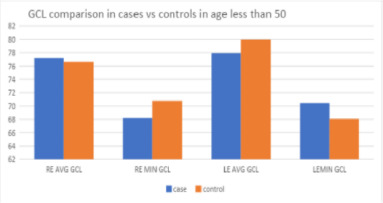
RE Avg RNFL and LE Avg RNFL showed weak correlations with ALSFRS, indicated by Pearson Correlation coefficients of 0.073 and -0.026, respectively. The p-values (0.637 and 0.86) suggested that these correlations were not statistically significant. RE Avg GCL and LE Avg GCL, on the other hand, exhibited moderate positive correlations with ALSFRS scores, with correlation coefficients of 0.337 (RE) and 0.389 (LE). These correlations were statistically significant, as indicated by p-values of 0.021 and 0.006, respectively, suggesting a substantial association between GCL thickness and ALS functional outcomes.

DISCUSSION

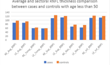
All patients in our study were clinically diagnosed cases of ALS, as per the El Escorial criteria. Age group-wise analysis showed statistically significant thinning overall as well as quadrant-wise RNFL parameters in patients less than 50 years compared to age-matched controls, indicating that the pathological process occurring in larger motor neurons in ALS might also be happening in smaller sensory neurons of the retina, causing thinning, which was not due to age-related process. Although GCIPL thinning was occurring in our cases, though statistically not significant compared to control, the significant positive correlation observed between GCIPL and ALS functional outcome and between RNFL and GCIPL measurements highlighted the fact that though the axonal degeneration in retinal neurons might not be translating to the same extent in ganglion cells in ALS, the subtle thinning of GCIPL correlated strongly with functional disability in patients with ALS, implying better functional scores with higher values of GCIPL parameters.

CONCLUSION

In summary, GCL measurements in both eyes showed a notable relationship with ALSFRS, whereas RNFL did not appear to correlate significantly.

摘要

目的

本研究旨在评估诊断为肌萎缩侧索硬化症(ALS)患者的各种光学相干断层扫描(OCT)参数。

方法

使用斯内伦视力表评估最佳矫正视力(BCVA),并进行主观验光以获得远视力和近视力的BCVA。采用戈德曼压平眼压计测量眼压(IOP)。使用90D透镜进行立体眼底检查以评估视神经和视网膜的状态,排除任何眼部病变。然后对患者进行OCT扫描以测量视神经乳头和黄斑参数。使用CIRRUS™ HD OCT(500 - 21822)(版本8.0.0.518)(卡尔蔡司医疗技术公司,美国加利福尼亚州都柏林)进行光学相干断层扫描。手动将分析区域居中,并确认每次扫描均无分割错误。

结果

右眼平均视网膜神经纤维层(RNFL)和左眼平均RNFL与肌萎缩侧索硬化功能评分量表(ALSFRS)呈弱相关性,皮尔逊相关系数分别为0.073和 - 0.026。p值(0.637和0.86)表明这些相关性无统计学意义。另一方面,右眼平均神经节细胞层(GCL)和左眼平均GCL与ALSFRS评分呈中度正相关,相关系数分别为0.337(右眼)和0.389(左眼)。这些相关性具有统计学意义,p值分别为0.021和0.006,表明GCL厚度与ALS功能结果之间存在显著关联。

讨论

根据埃尔埃斯科里亚尔标准,我们研究中的所有患者均为临床诊断的ALS病例。按年龄组分析显示,与年龄匹配的对照组相比,小于50岁的患者总体上以及象限性RNFL参数在统计学上有显著变薄,这表明ALS中较大运动神经元发生的病理过程可能也发生在视网膜较小的感觉神经元中,导致变薄,这并非与年龄相关的过程。尽管在我们的病例中发生了神经节细胞内层(GCIPL)变薄,与对照组相比虽无统计学意义,但在GCIPL与ALS功能结果之间以及RNFL与GCIPL测量之间观察到的显著正相关突出了这样一个事实,即尽管视网膜神经元中的轴突退化在ALS的神经节细胞中可能没有同等程度的表现,但GCIPL的细微变薄与ALS患者的功能残疾密切相关,这意味着GCIPL参数值越高,功能评分越好。

结论

总之,双眼的GCL测量与ALSFRS显示出显著关系,而RNFL似乎没有显著相关性。

https://cdn.ncbi.nlm.nih.gov/pmc/blobs/92e3/12277982/9e78b973f2fd/RomJOphthalmol-69-200-g004.jpg
https://cdn.ncbi.nlm.nih.gov/pmc/blobs/92e3/12277982/2a8847b24348/RomJOphthalmol-69-200-g001.jpg
https://cdn.ncbi.nlm.nih.gov/pmc/blobs/92e3/12277982/5b268d6f5aa3/RomJOphthalmol-69-200-g002.jpg
https://cdn.ncbi.nlm.nih.gov/pmc/blobs/92e3/12277982/9a1fd307fc1a/RomJOphthalmol-69-200-g003.jpg
https://cdn.ncbi.nlm.nih.gov/pmc/blobs/92e3/12277982/9e78b973f2fd/RomJOphthalmol-69-200-g004.jpg

相似文献

1
Ganglion Cell Layer Thickness as a Biomarker for Amyotrophic Lateral Sclerosis Functional Outcome: An OCT study.神经节细胞层厚度作为肌萎缩侧索硬化症功能预后的生物标志物:一项光学相干断层扫描研究。
Rom J Ophthalmol. 2025 Apr-Jun;69(2):200-207. doi: 10.22336/rjo.2025.32.
2
Regional retinal vulnerability in multiple sclerosis: integrating OCT, MRI, and clinical data for enhanced diagnosis and automated monitoring.多发性硬化症中的区域视网膜易损性:整合光学相干断层扫描(OCT)、磁共振成像(MRI)和临床数据以加强诊断和自动监测。
Rom J Morphol Embryol. 2025 Jan-Mar;66(1):119-130. doi: 10.47162/RJME.66.1.11.
3
Optic nerve head and fibre layer imaging for diagnosing glaucoma.用于诊断青光眼的视神经乳头和纤维层成像。
Cochrane Database Syst Rev. 2015 Nov 30;2015(11):CD008803. doi: 10.1002/14651858.CD008803.pub2.
4
Retinal layer segmentation in multiple sclerosis: a systematic review and meta-analysis.多发性硬化症中的视网膜层分割:系统评价和荟萃分析。
Lancet Neurol. 2017 Oct;16(10):797-812. doi: 10.1016/S1474-4422(17)30278-8. Epub 2017 Sep 12.
5
Macular versus Retinal Nerve Fiber Layer Parameters for Diagnosing Manifest Glaucoma: A Systematic Review of Diagnostic Accuracy Studies.黄斑与视网膜神经纤维层参数在诊断显性青光眼的应用:诊断准确性研究的系统评价。
Ophthalmology. 2016 May;123(5):939-49. doi: 10.1016/j.ophtha.2015.12.041. Epub 2016 Feb 15.
6
Optical coherence tomography (OCT) for detection of macular oedema in patients with diabetic retinopathy.光学相干断层扫描(OCT)用于检测糖尿病视网膜病变患者的黄斑水肿。
Cochrane Database Syst Rev. 2015 Jan 7;1(1):CD008081. doi: 10.1002/14651858.CD008081.pub3.
7
Alterations of the Ganglion Cell Complex in Age-Related Macular Degeneration: An Amish Eye Study Analysis.年龄相关性黄斑变性中神经节细胞复合体的改变:一项门诺派眼部研究分析。
Am J Ophthalmol. 2024 Sep;265:80-87. doi: 10.1016/j.ajo.2024.04.024. Epub 2024 Apr 26.
8
Associations of refractive errors and retinal changes measured by optical coherence tomography: A systematic review and meta-analysis.屈光不正与光学相干断层扫描测量的视网膜变化的相关性:系统评价和荟萃分析。
Surv Ophthalmol. 2022 Mar-Apr;67(2):591-607. doi: 10.1016/j.survophthal.2021.07.007. Epub 2021 Jul 31.
9
The Value of Optical Coherence Tomography in Patients with Pituitary Adenoma and Its Association with Clinical Features: A Pilot Study.光学相干断层扫描在垂体腺瘤患者中的价值及其与临床特征的关联:一项初步研究。
J Clin Med. 2025 Jun 17;14(12):4318. doi: 10.3390/jcm14124318.
10
Optical coherence tomography and angiography reveal early retinal alterations in pediatric-onset multiple sclerosis.光学相干断层扫描和血管造影显示儿童期多发性硬化症患者早期视网膜改变。
Eur J Pediatr. 2025 Jun 6;184(7):398. doi: 10.1007/s00431-025-06234-1.

本文引用的文献

1
Amyotrophic lateral sclerosis.肌萎缩侧索硬化症。
Nat Rev Dis Primers. 2017 Oct 5;3:17071. doi: 10.1038/nrdp.2017.71.
2
Amyotrophic Lateral Sclerosis.肌萎缩侧索硬化症
N Engl J Med. 2017 Jul 13;377(2):162-172. doi: 10.1056/NEJMra1603471.
3
Ophthalmic Manifestations of Amyotrophic Lateral Sclerosis (An American Ophthalmological Society Thesis).肌萎缩侧索硬化症的眼部表现(美国眼科学会论文)
Trans Am Ophthalmol Soc. 2015;113:T12.
4
Subtle retinal pathology in amyotrophic lateral sclerosis.肌萎缩侧索硬化症的细微视网膜病理学。
Ann Clin Transl Neurol. 2014 Apr;1(4):290-7. doi: 10.1002/acn3.46. Epub 2014 Mar 11.
5
Clinicopathologic report of ocular involvement in ALS patients with C9orf72 mutation.携带C9orf72突变的肌萎缩侧索硬化症患者眼部受累的临床病理报告。
Amyotroph Lateral Scler Frontotemporal Degener. 2014 Dec;15(7-8):569-80. doi: 10.3109/21678421.2014.951941. Epub 2014 Oct 16.
6
Detection of Retinal Nerve Fiber Layer Defects in Alzheimer's Disease Using SD-OCT.使用 SD-OCT 检测阿尔茨海默病的视网膜神经纤维层缺陷。
Front Psychiatry. 2014 Feb 25;5:22. doi: 10.3389/fpsyt.2014.00022. eCollection 2014.
7
Optical coherence tomography does not support optic nerve involvement in amyotrophic lateral sclerosis.光学相干断层扫描不支持在肌萎缩侧索硬化症中视神经受累。
Eur J Neurol. 2013 Aug;20(8):1170-6. doi: 10.1111/ene.12146. Epub 2013 Apr 14.
8
Retinal damage in multiple sclerosis disease subtypes measured by high-resolution optical coherence tomography.通过高分辨率光学相干断层扫描测量的多发性硬化症疾病亚型中的视网膜损伤。
Mult Scler Int. 2012;2012:530305. doi: 10.1155/2012/530305. Epub 2012 Jul 25.
9
Optic nerve head quantification in idiopathic intracranial hypertension by spectral domain OCT.特发性颅内高压的视神经头定量分析:频域光学相干断层扫描。
PLoS One. 2012;7(5):e36965. doi: 10.1371/journal.pone.0036965. Epub 2012 May 15.
10
Optical coherence tomography in parkinsonian syndromes.帕金森综合征的光学相干断层扫描。
PLoS One. 2012;7(4):e34891. doi: 10.1371/journal.pone.0034891. Epub 2012 Apr 13.