• 文献检索
  • 文档翻译
  • 深度研究
  • 学术资讯
  • Suppr Zotero 插件Zotero 插件
  • 邀请有礼
  • 套餐&价格
  • 历史记录
应用&插件
Suppr Zotero 插件Zotero 插件浏览器插件Mac 客户端Windows 客户端微信小程序
定价
高级版会员购买积分包购买API积分包
服务
文献检索文档翻译深度研究API 文档MCP 服务
关于我们
关于 Suppr公司介绍联系我们用户协议隐私条款
关注我们

Suppr 超能文献

核心技术专利:CN118964589B侵权必究
粤ICP备2023148730 号-1Suppr @ 2026

文献检索

告别复杂PubMed语法,用中文像聊天一样搜索,搜遍4000万医学文献。AI智能推荐,让科研检索更轻松。

立即免费搜索

文件翻译

保留排版,准确专业,支持PDF/Word/PPT等文件格式,支持 12+语言互译。

免费翻译文档

深度研究

AI帮你快速写综述,25分钟生成高质量综述,智能提取关键信息,辅助科研写作。

立即免费体验

遗传性视网膜疾病中的眼底自发荧光:综述

Fundus Autofluorescence in Inherited Retinal Disease: A Review.

作者信息

Oh Jin Kyun, Moussa Omar, Lam Byron L, Sengillo Jesse D

机构信息

Department of Ophthalmology, Vagelos College of Physicians and Surgeons, Columbia University Irving Medical Center, New York, NY 10032, USA.

Department of Ophthalmology, Bascom Palmer Eye Institute, School of Medicine, University of Miami Miller, Miami, FL 33136, USA.

出版信息

Cells. 2025 Jul 16;14(14):1092. doi: 10.3390/cells14141092.

DOI:10.3390/cells14141092
PMID:40710345
原文链接:https://pmc.ncbi.nlm.nih.gov/articles/PMC12293359/
Abstract

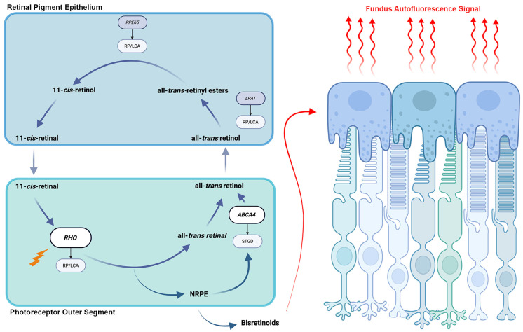
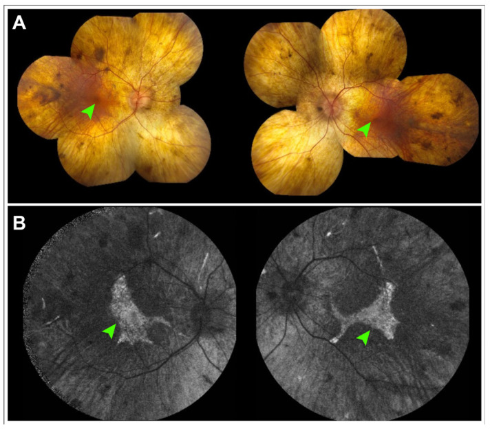
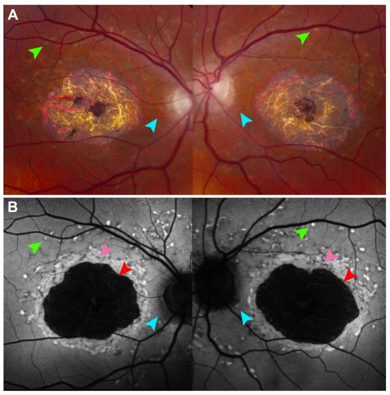
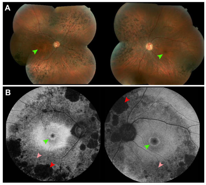
Fundus autofluorescence (FAF) is a non-invasive retinal imaging technique that helps visualize naturally occurring fluorophores, such as lipofuscin, and provides valuable insight into retinal diseases-particularly inherited retinal diseases (IRDs). FAF is especially useful in detecting subclinical or early-stage IRDs and in monitoring disease progression over time. In Stargardt disease, areas of decreased autofluorescence correlate with disease progression and have been proposed as a biomarker for future clinical trials. FAF can also help differentiate Stargardt disease from other macular dystrophies. In retinitis pigmentosa, hyperautofluorescent rings are a common feature on FAF and serve as an important marker for disease monitoring, especially as changes align with those seen on other imaging modalities. FAF is valuable in tracking progression of choroideremia and may help identify disease carrier status. FAF has also improved the characterization of mitochondrial retinopathies such as maternally inherited diabetes and deafness. As a rapid and widely accessible imaging modality, FAF plays a critical role in both diagnosis and longitudinal care of patients with IRDs.

摘要

眼底自发荧光(FAF)是一种非侵入性视网膜成像技术,有助于可视化天然存在的荧光团,如脂褐素,并为视网膜疾病,尤其是遗传性视网膜疾病(IRD)提供有价值的见解。FAF在检测亚临床或早期IRD以及监测疾病随时间的进展方面特别有用。在斯塔加特病中,自发荧光降低的区域与疾病进展相关,并已被提议作为未来临床试验的生物标志物。FAF还可以帮助区分斯塔加特病与其他黄斑营养不良。在色素性视网膜炎中,高自发荧光环是FAF上的常见特征,是疾病监测的重要标志物,尤其是当变化与其他成像方式所见的变化一致时。FAF在追踪脉络膜视网膜炎的进展方面很有价值,可能有助于确定疾病携带者状态。FAF还改善了对线粒体视网膜病变(如母系遗传的糖尿病和耳聋)的特征描述。作为一种快速且广泛可用的成像方式,FAF在IRD患者的诊断和长期护理中都起着关键作用。

https://cdn.ncbi.nlm.nih.gov/pmc/blobs/9541/12293359/6364d83380a7/cells-14-01092-g004.jpg
https://cdn.ncbi.nlm.nih.gov/pmc/blobs/9541/12293359/d35df8f9d5d1/cells-14-01092-g001.jpg
https://cdn.ncbi.nlm.nih.gov/pmc/blobs/9541/12293359/ee867b41528e/cells-14-01092-g002.jpg
https://cdn.ncbi.nlm.nih.gov/pmc/blobs/9541/12293359/31c3e066bbc9/cells-14-01092-g003.jpg
https://cdn.ncbi.nlm.nih.gov/pmc/blobs/9541/12293359/6364d83380a7/cells-14-01092-g004.jpg

相似文献

1
Fundus Autofluorescence in Inherited Retinal Disease: A Review.遗传性视网膜疾病中的眼底自发荧光:综述
Cells. 2025 Jul 16;14(14):1092. doi: 10.3390/cells14141092.
2
Accuracy of fundus autofluorescence imaging for the diagnosis and monitoring of retinal conditions: a systematic review.眼底自发荧光成像在视网膜疾病诊断和监测中的准确性:一项系统评价
Health Technol Assess. 2016 Apr;20(31):1-108. doi: 10.3310/hta20310.
3
Blue Light and Green Light Fundus Autofluorescence, Complementary to Optical Coherence Tomography, in Age-Related Macular Degeneration Evaluation.蓝光和绿光眼底自发荧光在年龄相关性黄斑变性评估中对光学相干断层扫描的补充作用
Diagnostics (Basel). 2025 Jul 2;15(13):1688. doi: 10.3390/diagnostics15131688.
4
The Black Book of Psychotropic Dosing and Monitoring.《精神药物剂量与监测黑皮书》
Psychopharmacol Bull. 2024 Jul 8;54(3):8-59.
5
Management of urinary stones by experts in stone disease (ESD 2025).结石病专家对尿路结石的管理(2025年结石病专家共识)
Arch Ital Urol Androl. 2025 Jun 30;97(2):14085. doi: 10.4081/aiua.2025.14085.
6
Retinopathy in Mucopolysaccharidoses.黏多糖贮积症中的视网膜病变
Ophthalmology. 2025 Apr;132(4):461-475. doi: 10.1016/j.ophtha.2024.11.013. Epub 2024 Nov 14.
7
Fundus autofluorescence imaging: systematic review of test accuracy for the diagnosis and monitoring of retinal conditions.眼底自发荧光成像:视网膜疾病诊断与监测的检测准确性系统评价
Eye (Lond). 2017 Jul;31(7):995-1007. doi: 10.1038/eye.2017.19. Epub 2017 Mar 10.
8
Short-Term Memory Impairment短期记忆障碍
9
Progression Rate of Macular Retinal Pigment Epithelium Atrophy in Geographic Atrophy and Selected Inherited Retinal Dystrophies. A Systematic Review and Meta-Analysis.地图样萎缩和特定遗传性视网膜营养不良中黄斑视网膜色素上皮萎缩的进展率。一项系统评价和荟萃分析。
Am J Ophthalmol. 2025 Jan;269:30-48. doi: 10.1016/j.ajo.2024.07.035. Epub 2024 Aug 15.
10
Optical coherence tomography (OCT) for detection of macular oedema in patients with diabetic retinopathy.光学相干断层扫描(OCT)用于检测糖尿病视网膜病变患者的黄斑水肿。
Cochrane Database Syst Rev. 2015 Jan 7;1(1):CD008081. doi: 10.1002/14651858.CD008081.pub3.

本文引用的文献

1
Multimodal Imaging and Dark-Adapted Chromatic Perimetry in BEST1 Vitelliform Macular Dystrophy: Identification of Outcome Measurements.最佳1型卵黄样黄斑营养不良的多模态成像与暗适应色觉视野检查:结局测量指标的确定
Ophthalmol Sci. 2025 May 6;5(5):100823. doi: 10.1016/j.xops.2025.100823. eCollection 2025 Sep-Oct.
2
Ellipsoid Zone Loss as an Outcome Measure in MAK-Associated Retinitis Pigmentosa.椭球区丢失作为常染色体显性遗传性视网膜色素变性相关的一种结局指标
Am J Ophthalmol. 2025 Jun 25;278:282-291. doi: 10.1016/j.ajo.2025.06.031.
3
Near-Infrared Autofluorescence: Early Detection of Retinal Pigment Epithelial Alterations in Inherited Retinal Dystrophies.
近红外自发荧光:遗传性视网膜营养不良中视网膜色素上皮改变的早期检测
J Clin Med. 2024 Nov 15;13(22):6886. doi: 10.3390/jcm13226886.
4
Retinal Patterns and the Role of Autofluorescence in Choroideremia.视网膜模式与脉络膜黑色素瘤中自发荧光的作用。
Genes (Basel). 2024 Nov 14;15(11):1471. doi: 10.3390/genes15111471.
5
XOLARIS: A 24-Month, Prospective, Natural History Study of 201 Participants with -Associated X-Linked Retinitis Pigmentosa.XOLARIS:一项针对201名与X连锁视网膜色素变性相关患者的为期24个月的前瞻性自然史研究。
Ophthalmol Sci. 2024 Aug 13;5(1):100595. doi: 10.1016/j.xops.2024.100595. eCollection 2025 Jan-Feb.
6
Endpoints and Design for Clinical Trials in USH2A-Related Retinal Degeneration: Results and Recommendations From the RUSH2A Natural History Study.USH2A 相关视网膜变性临床试验的终点和设计:RUSH2A 自然史研究的结果和建议。
Transl Vis Sci Technol. 2024 Oct 1;13(10):15. doi: 10.1167/tvst.13.10.15.
7
Current and Future Directions in Developing Effective Treatments for PRPH2-Associated Retinal Diseases: A Workshop Report.开发有效治疗 PRPH2 相关视网膜疾病的当前和未来方向:研讨会报告。
Transl Vis Sci Technol. 2024 Oct 1;13(10):16. doi: 10.1167/tvst.13.10.16.
8
PDE6A-Associated Retinitis Pigmentosa, Clinical Characteristics, Genetics, and Natural History.与磷酸二酯酶6A相关的色素性视网膜炎:临床特征、遗传学及自然病史
Ophthalmol Retina. 2025 Mar;9(3):278-287. doi: 10.1016/j.oret.2024.08.018. Epub 2024 Aug 31.
9
CDH23-Associated Usher Syndrome: Clinical Features, Retinal Imaging, and Natural History.与CDH23相关的Usher综合征:临床特征、视网膜成像及自然病史
Invest Ophthalmol Vis Sci. 2024 Jul 1;65(8):27. doi: 10.1167/iovs.65.8.27.
10
Mitochondrial retinopathies and optic neuropathies: The impact of retinal imaging on modern understanding of pathogenesis, diagnosis, and management.线粒体性视网膜病和视神经病变:视网膜成像对发病机制、诊断和治疗的现代认识的影响。
Prog Retin Eye Res. 2024 Jul;101:101264. doi: 10.1016/j.preteyeres.2024.101264. Epub 2024 May 3.