Maldonado Andres A, Broski Stephen M, Marek Tomas, Cristóbal Lara, Spinner Robert J
Department of Neurologic Surgery, Mayo Clinic, Gonda 8-214, Rochester, MN, 55905, USA.
Department of Plastic and Reconstructive Surgery, Hospital Universitario de Getafe, Madrid, Spain.
Acta Neurochir (Wien). 2025 Jul 29;167(1):207. doi: 10.1007/s00701-025-06615-3.
Perineural spread (PNS) of breast carcinoma to the brachial plexus is rare. This study investigates the radiologic features supporting the medial and lateral pectoral nerves (MPN and LPN, respectively) as pathways for PNS of breast cancer to the brachial plexus.
We reviewed 19 patients with biopsy-proven PNS of breast carcinoma to the brachial plexus. All available MRI and F-FDG PET/CT studies were re-evaluated by a musculoskeletal radiologist with expertise in PNS. Imaging features of interest included pectoralis major and minor muscle MRI signal abnormality, abnormal FDG activity, and atrophy; FDG avidity within or along the course of the pectoral nerves; and extent of brachial plexus involvement on MRI and F-FDG PET/CT. Demographic and clinical data were also collected.
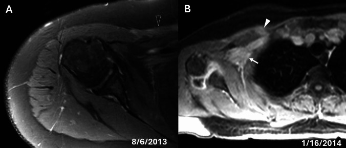
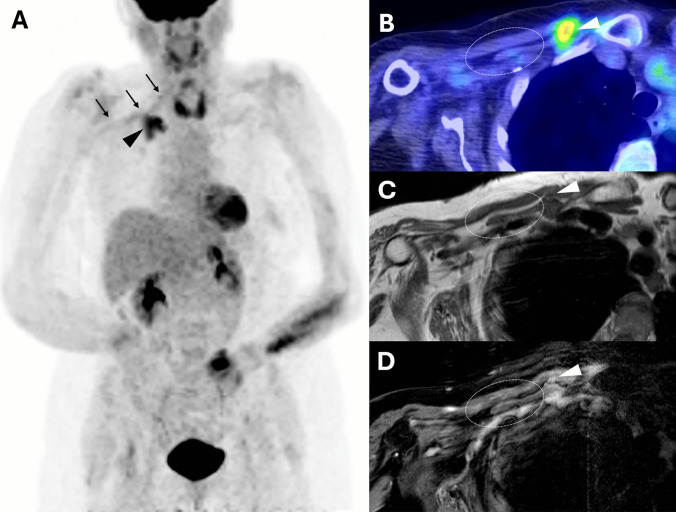
All 19 patients had MRI and F-FDG PET/CT scans. Six patients showed clear radiologic evidence of PNS via the pectoral nerves. All six patients demonstrated abnormal MRI signal or enhancement in both the pectoralis major and minor muscles and increased FDG uptake was present in the pectoralis major in 4/6 patients and pectoralis minor in 5/6 patients. Five patients demonstrated atrophy of both the pectoralis major and minor muscles. Increased FDG uptake was noted along the LPN in five patients and the MPN in four. All exhibited brachial plexus enhancement on MRI and increased FDG uptake on PET/CT, supporting contiguous spread from the pectoral nerves.
This study provides radiologic support for the MPN and LPN as a potential pathway for PNS of breast cancer to the brachial plexus. Pectoralis major/minor muscle atrophy, abnormal MRI signal or enhancement, and increased FDG activity within the pectoral muscles and/or along the pectoral nerves may serve as early, non-invasive imaging markers of this process, with potential implications for diagnosis and management.
乳腺癌向臂丛神经的神经周围扩散(PNS)较为罕见。本研究调查支持胸内侧神经和胸外侧神经(分别为MPN和LPN)作为乳腺癌向臂丛神经PNS途径的放射学特征。
我们回顾了19例经活检证实乳腺癌向臂丛神经发生PNS的患者。所有可用的MRI和F-FDG PET/CT研究均由一位在PNS方面有专长的肌肉骨骼放射科医生重新评估。感兴趣的影像特征包括胸大肌和胸小肌的MRI信号异常、FDG活性异常和萎缩;胸神经内或沿胸神经走行的FDG摄取;以及MRI和F-FDG PET/CT上臂丛神经受累的范围。还收集了人口统计学和临床数据。
所有19例患者均进行了MRI和F-FDG PET/CT扫描。6例患者显示出通过胸神经发生PNS的明确放射学证据。所有6例患者胸大肌和胸小肌均表现出异常的MRI信号或强化,4/6例患者胸大肌FDG摄取增加,5/6例患者胸小肌FDG摄取增加。5例患者胸大肌和胸小肌均出现萎缩。5例患者LPN沿线FDG摄取增加,4例患者MPN沿线FDG摄取增加。所有患者MRI上均显示臂丛神经强化,PET/CT上FDG摄取增加,支持从胸神经的连续性扩散。
本研究为MPN和LPN作为乳腺癌向臂丛神经PNS的潜在途径提供了放射学支持。胸大肌/胸小肌萎缩、MRI信号异常或强化以及胸肌内和/或胸神经沿线FDG活性增加可能作为这一过程的早期非侵入性影像标志物,对诊断和管理具有潜在意义。