Üremiş Muhammed Mehdi, Üremiş Nuray, Ceylan Mustafa, Türköz Yusuf
İnönü University Faculty of Medicine, Department of Medical Biochemistry, Malatya, Türkiye.
Gaziosmanpaşa University Faculty of Arts and Sciences, Department of Chemistry, Tokat, Türkiye.
Turk J Pharm Sci. 2025 Aug 1;22(3):207-216. doi: 10.4274/tjps.galenos.2025.49840.
Benzothiazole compounds, characterized by their diverse biological and pharmacological properties, have emerged as promising molecules for suppressing cancer cell proliferation and invasion due to their antiproliferative attributes. Prior research from our laboratory revealed that 2-substituted benzothiazole compounds inhibit the proliferation of glioma and cervical cancer cells and induce apoptosis in pancreatic cancer cells. However, there is limited research on the effectiveness of benzothiazoles against hepatocellular carcinoma cells (HCC). This study sought to elucidate the anticancer potential of 2-substituted benzothiazole derivatives through their modulation of oxidative stress and inflammation mediators.
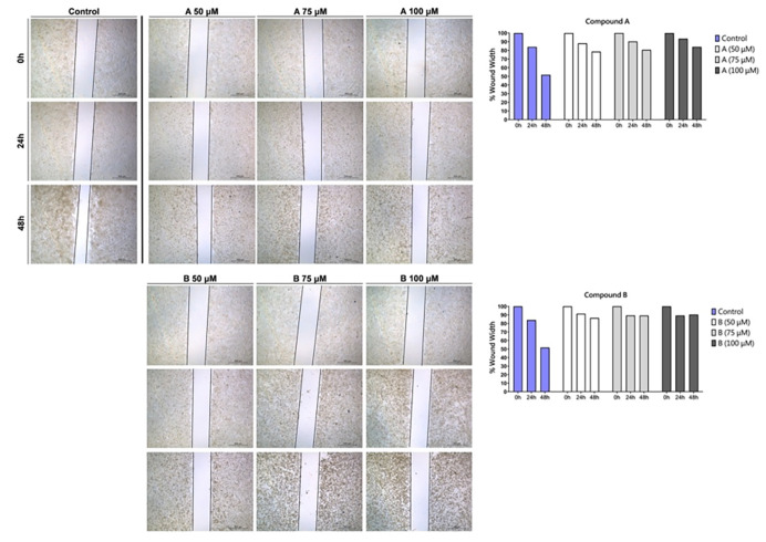
Antiproliferative effects of two-step synthesized 2-substituted benzothiazole derivatives were evaluated on HepG2 cells via MTT assay. Apoptosis induction was assessed using Annexin V/PI staining; cell cycle arrest effects were determined through cell cycle analysis; cell migration was examined via wound healing assay; and mitochondrial membrane damage was quantified using JC-1 staining. Spectrophotometric measurements of total antioxidant status (TAS), total oxidant status, superoxide dismutase (SOD), total thiol, and native thiol levels were used to assess cellular redox status. Expression of nuclear factor kappa B (NF-κB), an inflammatory marker, was assessed by western blot, while inflammation-related cyclooxygenase-2 (COX-2) and inducible nitric oxide synthase levels were measured using ELISA.
This investigation unveiled benzothiazole derivatives' antiproliferative and cytotoxic properties against HepG2 cells (IC values of 56.98 μM and 59.17 μM at 24 h, and 38.54 μM 29.63 at 48 h). The synthesized compounds exhibited the ability to suppress cell migration and induce apoptosis, mediated by mitochondrial membrane potential loss (wound‑closure rates of 84.0 and 90.4% vs. 51.7% control at 48 h, apoptosis rates of 10.70% and 45.22% vs. 1.02% control). Furthermore, these derivatives reduced SOD activity (A and B at 100 μM p<0.001), TAS levels (A and B at 100 μM, < 0.05, < 0.001), and dynamic disulfide content. Notably, a decrease in NF-κB protein levels, closely associated with inflammation, was observed, along with a subsequent reduction in downstream effectors COX-2 (A and B at 100 μM, p<0.001) and iNOS (A and B at 100 μM, < 0.001).
The findings of this study underscore the antiproliferative effects of benzothiazole derivatives in human HCCs, coupled with their anti-inflammatory potential by diminishing NF-κB levels.
苯并噻唑化合物具有多种生物学和药理学特性,由于其抗增殖特性,已成为抑制癌细胞增殖和侵袭的有前景的分子。我们实验室先前的研究表明,2-取代苯并噻唑化合物可抑制胶质瘤和宫颈癌细胞的增殖,并诱导胰腺癌细胞凋亡。然而,关于苯并噻唑对肝癌细胞(HCC)有效性的研究有限。本研究旨在通过调节氧化应激和炎症介质来阐明2-取代苯并噻唑衍生物的抗癌潜力。
通过MTT法评估两步合成的2-取代苯并噻唑衍生物对HepG2细胞的抗增殖作用。使用Annexin V/PI染色评估凋亡诱导情况;通过细胞周期分析确定细胞周期阻滞作用;通过伤口愈合试验检测细胞迁移;使用JC-1染色定量线粒体膜损伤。通过分光光度法测量总抗氧化状态(TAS)、总氧化状态、超氧化物歧化酶(SOD)、总硫醇和天然硫醇水平来评估细胞氧化还原状态。通过蛋白质印迹法评估炎症标志物核因子κB(NF-κB)的表达,同时使用酶联免疫吸附测定法测量炎症相关的环氧化酶-2(COX-2)和诱导型一氧化氮合酶水平。
本研究揭示了苯并噻唑衍生物对HepG2细胞的抗增殖和细胞毒性特性(24小时时IC值分别为56.98 μM和59.17 μM,48小时时为38.54 μM和29.63 μM)。合成的化合物表现出抑制细胞迁移和诱导凋亡的能力,这是由线粒体膜电位丧失介导的(48小时时伤口闭合率分别为84.0%和90.4%,而对照为51.7%,凋亡率分别为10.70%和45.22%,而对照为1.02%)。此外,这些衍生物降低了SOD活性(100 μM时A和B,p<0.001)、TAS水平(100 μM时A和B,<0.05,<0.001)和动态二硫键含量。值得注意的是,观察到与炎症密切相关的NF-κB蛋白水平降低,随后下游效应物COX-2(100 μM时A和B,p<0.001)和诱导型一氧化氮合酶(100 μM时A和B,<0.001)也随之降低。
本研究结果强调了苯并噻唑衍生物在人肝癌中的抗增殖作用,以及通过降低NF-κB水平所具有的抗炎潜力。