Ren Xueting, Wu Jinpeng, Li Jing, Zhai Zhen, Li Xiang, Guan Feng, Wang Meng, Ma Xiaobin, Tan Zengqi, Kang Huafeng, Lin Shuai
The Comprehensive Breast Care Center, The Second Affiliated Hospital of Xi'an Jiaotong University, Xi'an, Shaanxi, China.
Key Laboratory of Resource Biology and Biotechnology in Western China, Ministry of Education, College of Life Sciences, Northwest University, Xi'an, Shaanxi, China.
Exp Hematol Oncol. 2025 Aug 4;14(1):102. doi: 10.1186/s40164-025-00693-w.
The abundance of PD-L1 on the surface of tumor cells is a critical factor in sensitizing these cells to T cell-mediated immune killing. While abnormal glycosylation of PD-L1 is known to influence its expression and function, the precise regulatory mechanisms remain unclear.
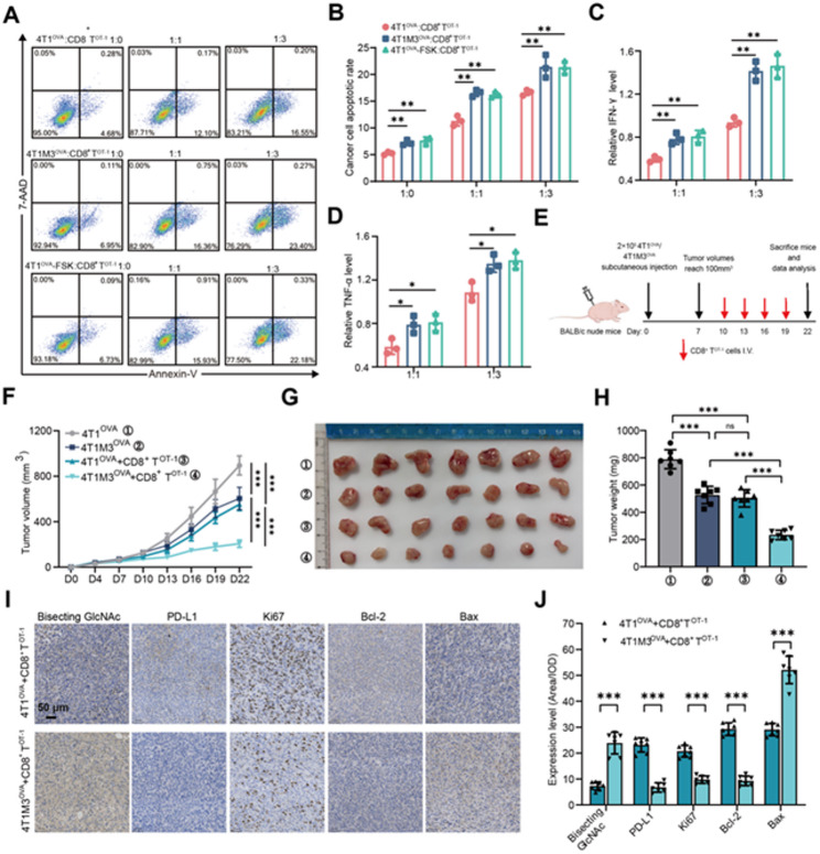
This study utilized bioinformatics analysis to explore the role of MGAT3, a key gene involved in the formation of the bisecting GlcNAc structure, in breast cancer (BC). Experimental approaches were employed to increase bisecting GlcNAc levels in BC cells, followed by assessments of PD-L1 expression, CD8 T cell-mediated cytotoxicity, extracellular vesicle (EV)-associated PD-L1, and PD-L1/PD-1 interaction. Additionally, forskolin, a bisecting GlcNAc agonist, was combined with anti-PD-L1 antibody to evaluate its antitumor effects in vivo.
MGAT3 was found to be expressed at low levels in BC tissues but positively correlated with CD8 T cell infiltration. Elevating bisecting GlcNAc levels in BC cells significantly enhanced the cytotoxic efficacy of CD8 T cells. High bisecting GlcNAc modification promoted PD-L1 degradation via the lysosomal pathway, reducing PD-L1 expression and its binding to PD-1. Furthermore, increased bisecting GlcNAc levels reduced PD-L1 in tumor cell-derived EVs, impairing the EVs' ability to block CD8 T cells and indirectly enhancing T cell cytotoxicity. The combined use of forskolin and anti-PD-L1 antibody significantly increased CD8 T cell abundance and activity, achieving a more effective antitumor response in vivo.
These findings demonstrate that enhancing bisecting GlcNAc modification in BC cells promotes PD-L1 degradation and inhibits its binding to PD-1, thereby boosting CD8 T cell-mediated cytotoxicity, providing a promising strategy for immune modulation in BC therapy.
肿瘤细胞表面PD-L1的丰度是使这些细胞对T细胞介导的免疫杀伤敏感的关键因素。虽然已知PD-L1的异常糖基化会影响其表达和功能,但确切的调控机制仍不清楚。
本研究利用生物信息学分析来探究参与平分型N-乙酰葡糖胺(GlcNAc)结构形成的关键基因MGAT3在乳腺癌(BC)中的作用。采用实验方法提高BC细胞中平分型GlcNAc的水平,随后评估PD-L1表达、CD8 T细胞介导的细胞毒性、细胞外囊泡(EV)相关的PD-L1以及PD-L1/PD-1相互作用。此外,将平分型GlcNAc激动剂福司可林与抗PD-L1抗体联合使用,以评估其在体内的抗肿瘤作用。
发现MGAT3在BC组织中低表达,但与CD8 T细胞浸润呈正相关。提高BC细胞中平分型GlcNAc的水平显著增强了CD8 T细胞的细胞毒性功效。高平分型GlcNAc修饰通过溶酶体途径促进PD-L1降解,降低PD-L1表达及其与PD-1的结合。此外,平分型GlcNAc水平的增加降低了肿瘤细胞衍生的EV中的PD-L1,损害了EV阻断CD8 T细胞的能力,并间接增强了T细胞的细胞毒性。福司可林与抗PD-L1抗体联合使用显著增加了CD8 T细胞的丰度和活性,在体内实现了更有效的抗肿瘤反应。
这些发现表明,增强BC细胞中的平分型GlcNAc修饰可促进PD-L1降解并抑制其与PD-1的结合,从而增强CD8 T细胞介导的细胞毒性,为BC治疗中的免疫调节提供了一种有前景的策略。