Rivas Dominic J L, Gassmann Joshua M, Goetz Jessica E, Aitken Holly D, Davison John C, Miller Aspen, Willey Michael C
University of Iowa Hospitals and Clinics, Department of Orthopedics and Rehabilitation, 200 Hawkins Dr., Iowa City, IA 52242, United States.
J Hip Preserv Surg. 2025 Jan 22;12(2):93-104. doi: 10.1093/jhps/hnaf001. eCollection 2025 Jul.
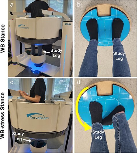
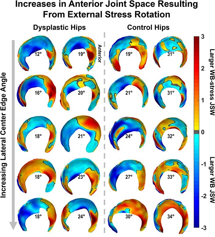
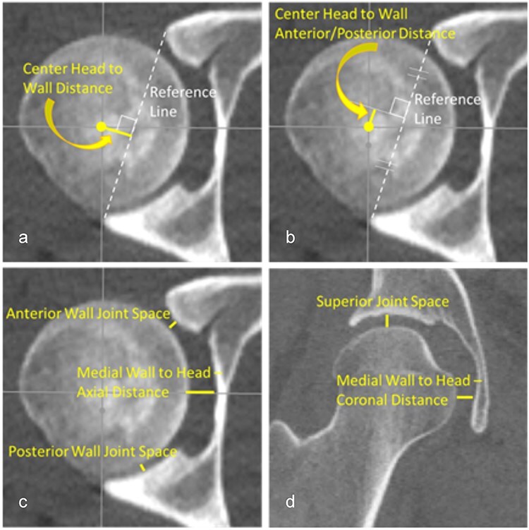
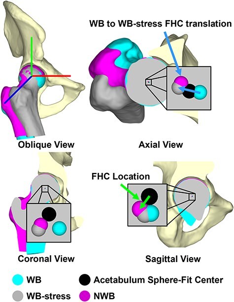
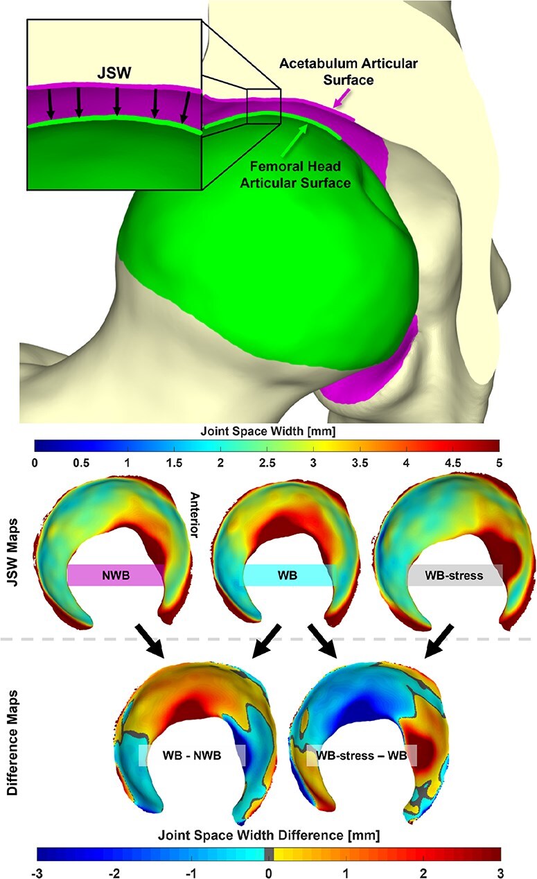
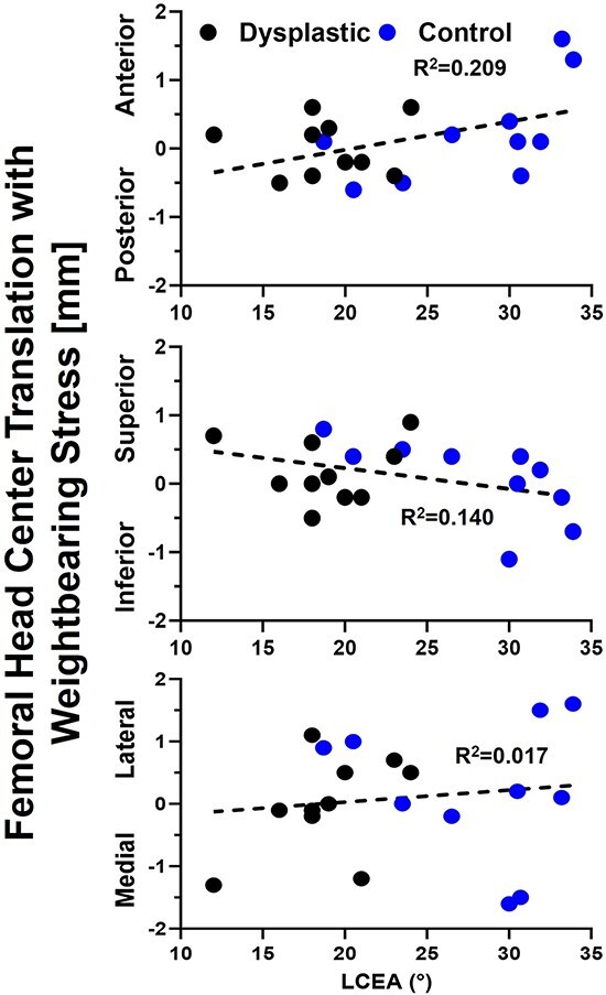
Hip dysplasia causes pathologic joint mechanics and can produce hip instability, leading to progressive joint degeneration and osteoarthritis. Weight-bearing computed tomography (WBCT) is an emerging technology that may enable quantification of femoral-acetabular displacement as an objective indicator of instability. To evaluate this potential, 10 patients indicated for periacetabular osteotomy to treat hip dysplasia and 10 healthy controls underwent two WBCT protocols. Participants were scanned in a neutral stance [weight-bearing (WB)] and again with the hip stressed in maximal external rotation (WB-stress), a position hypothesized to reproduce anterior instability. Clinical, nonweight-bearing computed tomography (CT) scans were available for patients with hip dysplasia. Congruency of the femoroacetabular joint space and position of the femoral head in the acetabulum were quantified via multiple 2D manual measurements and automated 3D measurements. There were no 2D measurements found to differ between the WB and WB-stress scans in either dysplastic (= .742-1.000) or control (= .203-1.000) hips. 3D translation of the femoral head center from WB to WB-stress averaged 1.3 ± 0.6 mm in the control hips, compared to 0.9 ± 0.4 mm in the dysplastic hips (= .096). 3D joint space width (JSW) was determined for both the control and dysplastic hips, with greater JSW found in control hips for both the WB (= .049) and WB-stress (= .003) scans. WBCT has the potential to better capture subtle femoral-acetabular displacement derived from both automated 3D and manual 2D measurements in static instability-prone joint orientations.
髋关节发育不良会导致病理性关节力学改变,并可能导致髋关节不稳定,进而引发渐进性关节退变和骨关节炎。负重计算机断层扫描(WBCT)是一项新兴技术,它或许能够量化股骨髋臼移位情况,作为不稳定的客观指标。为评估这一潜力,10例因髋臼周围截骨术治疗髋关节发育不良而入选的患者以及10名健康对照者接受了两种WBCT方案。参与者先在中立位(负重)进行扫描,然后在髋关节最大外旋位(负重-应力位)再次扫描,该位置被认为可重现前侧不稳定。患有髋关节发育不良的患者可获得临床非负重计算机断层扫描(CT)图像。通过多次二维手动测量和自动三维测量,对股骨髋臼关节间隙的一致性以及股骨头在髋臼中的位置进行了量化。在发育不良髋关节(=0.742 - 1.000)或对照髋关节(=0.203 - 1.000)中,二维测量结果在负重扫描和负重-应力扫描之间均未发现差异。对照髋关节中股骨头中心从负重位到负重-应力位的三维平移平均为1.3±0.6毫米,而发育不良髋关节中为0.9±0.4毫米(=0.096)。对对照髋关节和发育不良髋关节均测定了三维关节间隙宽度(JSW),在负重扫描(=0.049)和负重-应力扫描(=0.003)中,对照髋关节的JSW均更大。WBCT有潜力在静态易发生不稳定的关节方位中,通过自动三维测量和手动二维测量更好地捕捉细微的股骨髋臼移位情况。