文献检索文档翻译深度研究
Suppr Zotero 插件Zotero 插件
邀请有礼套餐&价格历史记录

新学期,新优惠

限时优惠:9月1日-9月22日

30天高级会员仅需29元

1天体验卡首发特惠仅需5.99元

了解详情
不再提醒
插件&应用
Suppr Zotero 插件Zotero 插件浏览器插件Mac 客户端Windows 客户端微信小程序
高级版
套餐订阅购买积分包
AI 工具
文献检索文档翻译深度研究
关于我们
关于 Suppr公司介绍联系我们用户协议隐私条款
关注我们

Suppr 超能文献

核心技术专利:CN118964589B侵权必究
粤ICP备2023148730 号-1Suppr @ 2025

基于影像组学的机器学习,利用脑转移瘤的T1增强磁共振成像鉴别肺鳞状细胞癌和腺癌

Radiomics-based machine learning for differentiating lung squamous cell carcinoma and adenocarcinoma using T1-enhanced MRI of brain metastases.

作者信息

Xia Xueming, Tan Qiaoyue, Du Wei, Gou Qiheng

机构信息

Division of Head & Neck Tumor Multimodality Treatment, Cancer Center, West China Hospital, Sichuan University, Chengdu, China.

Radiotherapy Physics and Technology Center, Cancer Center, West China Hospital, Sichuan University, Chengdu, China.

出版信息

Front Oncol. 2025 Jul 23;15:1599853. doi: 10.3389/fonc.2025.1599853. eCollection 2025.


DOI:10.3389/fonc.2025.1599853
PMID:40772029
原文链接:https://pmc.ncbi.nlm.nih.gov/articles/PMC12325419/
Abstract

OBJECTIVE: This study aims to develop and evaluate a radiomics-based machine learning model using T1-enhanced magnetic resonance imaging (MRI) features to differentiate between lung squamous cell carcinoma (SCC) and adenocarcinoma (AC) in patients with brain metastases (BMs). While prior studies have largely focused on primary lung tumors, our work uniquely targets metastatic brain lesions, which pose distinct diagnostic and therapeutic challenges. METHODS: 
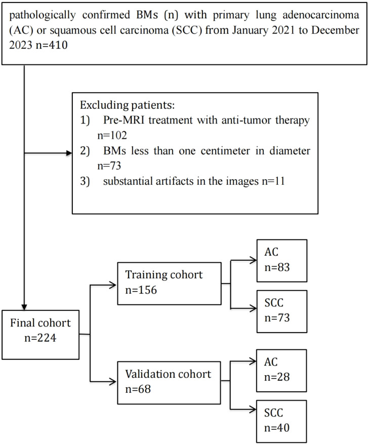
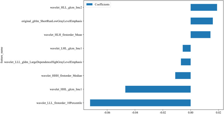
In this retrospective study, 173 patients with BMs from lung cancer were included, consisting of 88 with AC and 85 with SCC. MRI images were acquired using a standardized protocol, and 833 radiomic features were identified from the segmented lesions utilizing the PyRadiomics package. Feature selection was performed using a combination of univariate analysis, correlation analysis, and the least absolute shrinkage and selection operator (LASSO) regression. Ten machine learning classifiers were trained and validated utilizing the selected features. The performance of the classifier models was assessed through receiver operating characteristic (ROC) curves, and the area under the curve (AUC) was examined for analysis. RESULTS: 
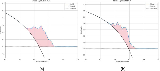
Ten classifier models were built on the basis of features derived from MRI. Among the ten classifier models, the LightGBM model performed the best. In the training dataset, the LightGBM classifier achieved an accuracy of 0.814, with a sensitivity of 0.726 and specificity of 0.896. The classifier's efficiency was validated on an independent testing dataset, where it maintained an accuracy of 0.779, with a sensitivity of 0.725 and specificity of 0.857. The AUC was 0.858 for the training dataset and 0.857 for the testing dataset. The model effectively distinguished between SCC and AC based on radiomic features, highlighting its potential for noninvasive non-small cell lung cancer (NSCLC) subtype classification. CONCLUSION: This research demonstrates the efficacy of a radiomics-based machine learning model in accurately classifying NSCLC subtypes from BMs, providing a valuable noninvasive tool for guiding personalized treatment strategies. Further validation on larger, multi-center datasets is crucial to verify these findings.

摘要

目的:本研究旨在开发并评估一种基于放射组学的机器学习模型,该模型利用T1增强磁共振成像(MRI)特征来区分脑转移瘤(BMs)患者的肺鳞状细胞癌(SCC)和腺癌(AC)。虽然先前的研究主要集中在原发性肺肿瘤上,但我们的工作独特地针对转移性脑病变,这些病变带来了独特的诊断和治疗挑战。 方法:在这项回顾性研究中,纳入了173例来自肺癌的脑转移瘤患者,其中88例为腺癌,85例为鳞状细胞癌。使用标准化方案采集MRI图像,并利用PyRadiomics软件包从分割的病变中识别出833个放射组学特征。采用单变量分析、相关性分析和最小绝对收缩和选择算子(LASSO)回归相结合的方法进行特征选择。利用选定的特征训练并验证了10个机器学习分类器。通过受试者操作特征(ROC)曲线评估分类器模型的性能,并检查曲线下面积(AUC)进行分析。 结果:基于MRI衍生的特征建立了10个分类器模型。在这10个分类器模型中,LightGBM模型表现最佳。在训练数据集中,LightGBM分类器的准确率为0.814,灵敏度为0.726,特异性为0.896。该分类器的效率在独立测试数据集中得到验证,其准确率保持在0.779,灵敏度为0.725,特异性为0.857。训练数据集的AUC为0.858,测试数据集的AUC为0.857。该模型基于放射组学特征有效地区分了鳞状细胞癌和腺癌,突出了其在非小细胞肺癌(NSCLC)亚型无创分类中的潜力。 结论:本研究证明了基于放射组学的机器学习模型在准确分类脑转移瘤的NSCLC亚型方面的有效性,为指导个性化治疗策略提供了一种有价值的无创工具。在更大的多中心数据集上进行进一步验证对于证实这些发现至关重要。

https://cdn.ncbi.nlm.nih.gov/pmc/blobs/4448/12325419/86a32c91df6c/fonc-15-1599853-g004.jpg
https://cdn.ncbi.nlm.nih.gov/pmc/blobs/4448/12325419/134c54936403/fonc-15-1599853-g001.jpg
https://cdn.ncbi.nlm.nih.gov/pmc/blobs/4448/12325419/efbae9e74de3/fonc-15-1599853-g002.jpg
https://cdn.ncbi.nlm.nih.gov/pmc/blobs/4448/12325419/0c64bfd817be/fonc-15-1599853-g003.jpg
https://cdn.ncbi.nlm.nih.gov/pmc/blobs/4448/12325419/86a32c91df6c/fonc-15-1599853-g004.jpg

相似文献

[1]
Radiomics-based machine learning for differentiating lung squamous cell carcinoma and adenocarcinoma using T1-enhanced MRI of brain metastases.

Front Oncol. 2025-7-23

[2]
Machine learning-based radiomics for differentiating lung cancer subtypes in brain metastases using CE-T1WI.

Front Oncol. 2025-6-19

[3]
Assessment of prostate cancer aggressiveness through the combined analysis of prostate MRI and 2.5D deep learning models.

Front Oncol. 2025-6-30

[4]
Radiomics Nomogram Based on Optimal Volume of Interest Derived from High-Resolution CT for Preoperative Prediction of IASLC Grading in Clinical IA Lung Adenocarcinomas: A Multi-Center, Large-Population Study.

Technol Cancer Res Treat. 2024

[5]
Machine learning-based brain magnetic resonance imaging radiomics for identifying rapid eye movement sleep behavior disorder in Parkinson's disease patients.

BMC Med Imaging. 2025-7-1

[6]
Prediction of EGFR Mutations in Lung Adenocarcinoma via CT Images: A Comparative Study of Intratumoral and Peritumoral Radiomics, Deep Learning, and Fusion Models.

Acad Radiol. 2025-5-5

[7]
Identification of the pathological subtypes of lung cancer brain metastases with multiparametric MRI radiomics: A feasibility study.

Sci Rep. 2025-7-23

[8]
Development and validation of a machine learning model for predicting co-infection of in pediatric patients.

Transl Pediatr. 2025-6-27

[9]
Multilayer perceptron deep learning radiomics model based on Gd-BOPTA MRI to identify vessels encapsulating tumor clusters in hepatocellular carcinoma: a multi-center study.

Cancer Imaging. 2025-7-7

[10]
Leveraging radiomics and machine learning to differentiate radiation necrosis from recurrence in patients with brain metastases.

J Neurooncol. 2024-6

本文引用的文献

[1]
Utilizing Radiomics of Peri-Lesional Edema in T2-FLAIR Subtraction Digital Images to Distinguish High-Grade Glial Tumors From Brain Metastasis.

J Magn Reson Imaging. 2025-4

[2]
Histological Subtype Classification of Non-Small Cell Lung Cancer with Radiomics and 3D Convolutional Neural Networks.

J Imaging Inform Med. 2024-12

[3]
[[Applications] 10. Radiomics Researches for Cancer Treatment Using Python].

Nihon Hoshasen Gijutsu Gakkai Zasshi. 2024

[4]
Non-Small Cell Lung Cancer, Version 4.2024, NCCN Clinical Practice Guidelines in Oncology.

J Natl Compr Canc Netw. 2024-5

[5]
Reproducibility in Radiomics: A Comparison of Feature Extraction Methods and Two Independent Datasets.

Appl Sci (Basel). 2024-2-20

[6]
Multi-omics and artificial intelligence predict clinical outcomes of immunotherapy in non-small cell lung cancer patients.

Clin Exp Med. 2024-3-30

[7]
Multisequence MRI-based radiomics signature as potential biomarkers for differentiating KRAS mutations in non-small cell lung cancer with brain metastases.

Eur J Radiol Open. 2024-1-16

[8]
Immunotherapy for lung cancer.

Pathol Res Pract. 2024-2

[9]
A systematic review of brain metastases from lung cancer using magnetic resonance neuroimaging: Clinical and technical aspects.

J Med Radiat Sci. 2024-6

[10]
Cancer statistics, 2024.

CA Cancer J Clin. 2024

文献AI研究员

20分钟写一篇综述,助力文献阅读效率提升50倍

立即体验

用中文搜PubMed

大模型驱动的PubMed中文搜索引擎

马上搜索

推荐工具

医学文档翻译智能文献检索