Torres Zapata Luis Enrique, Maldonado Calderón Jose Luis, Aguilar Urrea Luis Fernando, Garcia Bailón Aldo Missael, Molgado Garza Víctor Manuel
Urology, Hospital Universitario Dr. José Eleuterio González, Universidad Autónoma de Nuevo León, Monterrey, MEX.
Cureus. 2025 Jul 6;17(7):e87377. doi: 10.7759/cureus.87377. eCollection 2025 Jul.
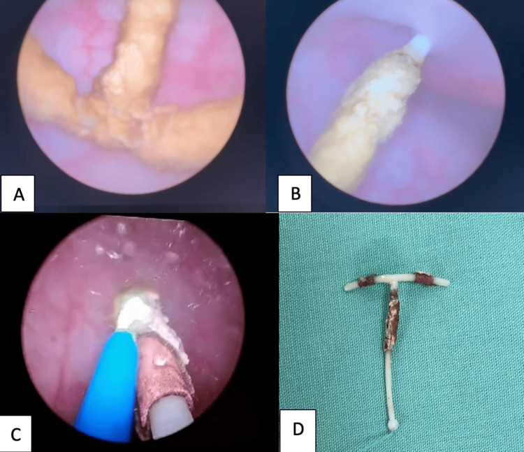
The intrauterine device (IUD) is a widely utilized method of contraception known for its high efficacy, safety profile, and long-term effectiveness. Despite its favorable characteristics, rare but potentially serious complications may occur, such as uterine perforation and device migration into adjacent pelvic or abdominal structures. One of the less frequent but clinically relevant complications is intravesical migration, where the device perforates the uterine wall and erodes into the urinary bladder. This can result in chronic lower urinary tract symptoms, recurrent urinary tract infections, hematuria, pelvic discomfort, and, in some cases, stone formation around the foreign body. We present the case of a 26-year-old female patient who developed recurrent urinary symptoms and intermittent hematuria three years after IUD placement. The device had not been visualized during gynecological follow-up and remained undetected during two full-term pregnancies. A noncontrast abdominal CT scan ultimately revealed a calcified IUD located within the urinary bladder. The patient underwent successful transurethral endoscopic removal using holmium:YAG laser lithotripsy in dusting mode to fragment the calcifications, followed by retrieval of the intact device with foreign body forceps. The procedure was completed without complications, and the patient reported full resolution of symptoms during follow-up. This case underscores the importance of considering IUD migration as a differential diagnosis in women presenting with unexplained urinary symptoms and a remote history of IUD use. It also demonstrates that holmium laser-assisted endoscopic management provides a safe, effective, and minimally invasive approach to remove encrusted or calcified intravesical IUDs, avoiding the need for open or laparoscopic surgery.
宫内节育器(IUD)是一种广泛应用的避孕方法,以其高效、安全和长期有效性而闻名。尽管其具有良好的特性,但仍可能发生罕见但潜在严重的并发症,如子宫穿孔和节育器迁移至邻近的盆腔或腹部结构。较不常见但临床上相关的并发症之一是膀胱内迁移,即节育器穿透子宫壁并侵蚀进入膀胱。这可能导致慢性下尿路症状、复发性尿路感染、血尿、盆腔不适,在某些情况下,还会在异物周围形成结石。我们报告一例26岁女性患者,她在放置IUD三年后出现复发性尿路症状和间歇性血尿。在妇科随访期间未发现该节育器,并且在两次足月妊娠期间均未被检测到。腹部非增强CT扫描最终显示一枚钙化的IUD位于膀胱内。患者接受了成功的经尿道内镜取出术,使用钬:YAG激光碎石机以粉末化模式粉碎钙化灶,随后用异物钳取出完整的节育器。手术完成且无并发症,患者在随访期间报告症状完全缓解。该病例强调了在出现不明原因尿路症状且有IUD使用史的女性中,将IUD迁移作为鉴别诊断的重要性。它还表明钬激光辅助内镜治疗为取出膀胱内结痂或钙化的IUD提供了一种安全、有效且微创的方法,避免了开放手术或腹腔镜手术的需要。