Li Li, Yang Yuting, Dai Fang, Deng Libin, Jiang Meixiu, He Chenjiang, Long Ting, Yang Kaiqiang, Yang Xinbo, Song Li
Center of Stomatology, The Second Affiliated Hospital, Jiangxi Medical College, Nanchang University, Nanchang, China.
JXHC Key Laboratory of Periodontology, (The Second Affiliated Hospital of Nanchang University), Nanchang, China.
J Transl Med. 2025 Aug 7;23(1):882. doi: 10.1186/s12967-025-06925-1.
Traditional techniques are limited in their ability to analyze the complex interaction mechanisms among multiple cell types within the periodontal microenvironment, thereby restricting the development of targeted therapies for periodontitis (PD). Utilizing multiomics technologies to investigate the interaction networks of key cell clusters can systematically uncover regulatory mechanisms and identify critical therapeutic targets.
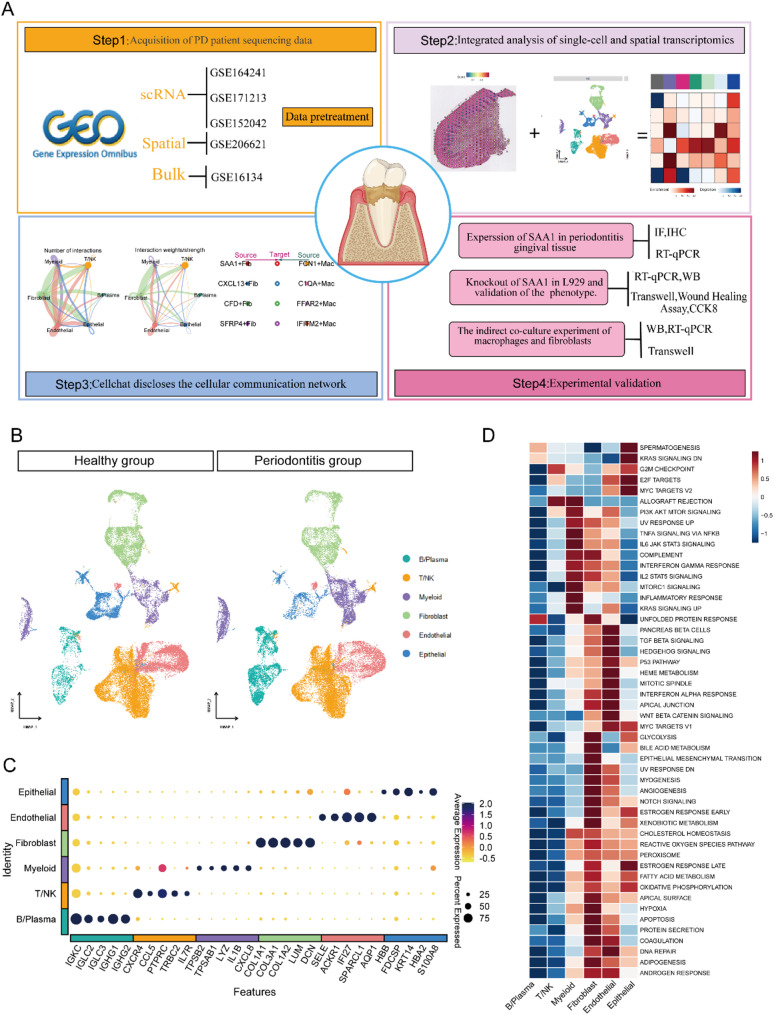
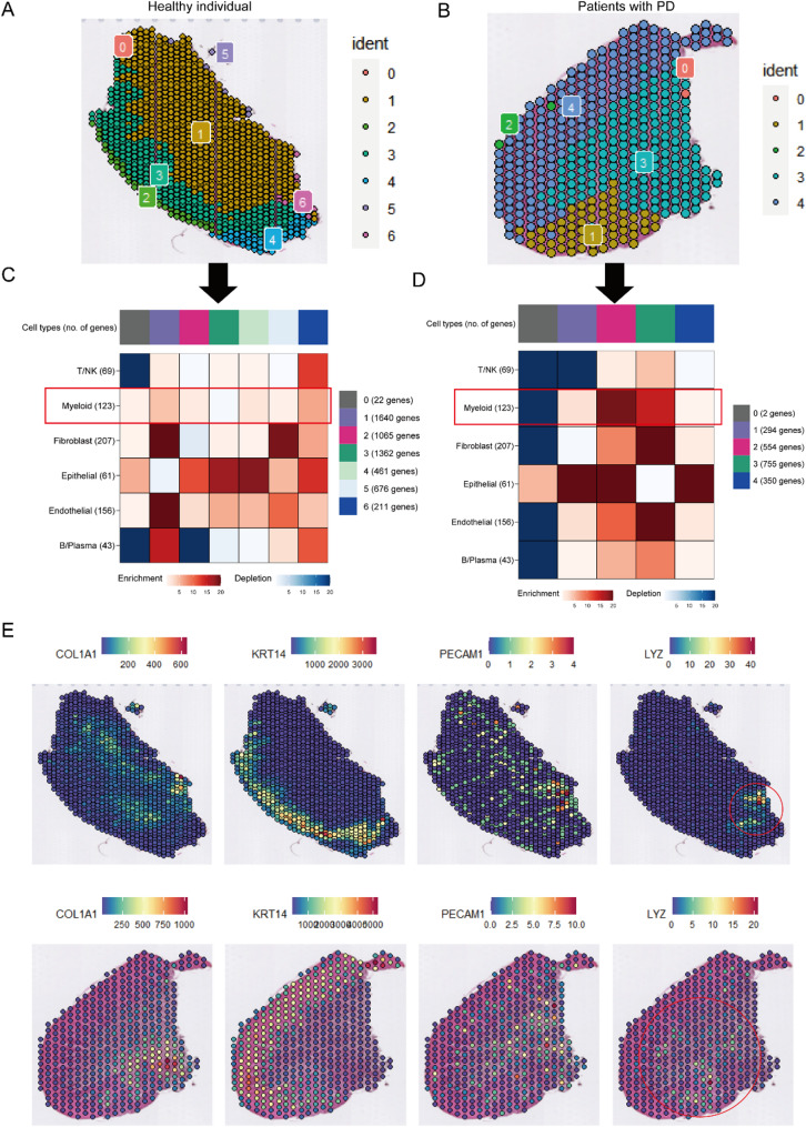
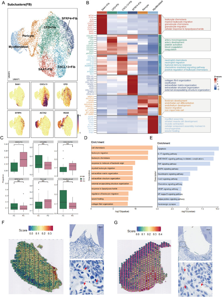
Through integrative analysis of single-cell RNA sequencing (scRNA-seq), spatial transcriptomics, and bulk transcriptome datasets from periodontal tissues, we systematically characterized the spatial architecture and intercellular communication networks within the inflammatory periodontal microenvironment, identifying a functionally serum amyloid A1 + fibroblasts (SAA1 + Fib) that critically drives disease progression. Combined bioinformatics and functional validations (in vitro and in vivo) revealed the proinflammatory role of SAA1 + Fib, demonstrating their unique transcriptional profile and mechanistic contributions to periodontal inflammation.
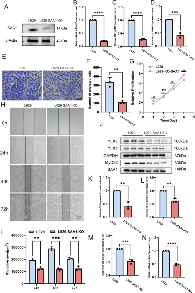
This study successfully constructed a single-cell transcriptome atlas comprising 65,979 periodontal tissue cells and identified an SAA1 + fibroblast subpopulation with key functions. Cell communication analysis revealed that this subpopulation mediates the infiltration of myeloid cells, such as macrophages, to the lesion site by secreting chemokine-related signaling molecules, including members of the SAA, CXCL, and CSF families. Animal experiments confirmed a significant increase in SAA1 expression levels in both the gingival tissue and peripheral blood of periodontitis model mice. Gene function studies indicated that SAA1 knockout resulted in reduced migration ability and enhanced proliferation activity of L929 cells, while significantly decreasing the secretion of inflammatory factors such as IL-6 and TNF-α. In a co-culture system of L929 cells and RAW264.7 cells, SAA1 knockout not only diminished the chemotactic effect of fibroblasts on macrophages but also suppressed the secretion of inflammatory factors and inhibited M1 polarization of macrophages. Mechanistic studies indicated that these effects were likely mediated by the suppression of NF-κB signaling pathway activity in RAW264.7 cells.
We elucidated the pro-inflammatory properties of SAA1 + Fib and their role in promoting macrophage infiltration, targeting SAA1 offers a new approach for the treatment of PD.
传统技术在分析牙周微环境中多种细胞类型之间复杂的相互作用机制方面存在局限性,从而限制了牙周炎(PD)靶向治疗的发展。利用多组学技术研究关键细胞簇的相互作用网络,可以系统地揭示调控机制并确定关键治疗靶点。
通过对来自牙周组织的单细胞RNA测序(scRNA-seq)、空间转录组学和批量转录组数据集进行综合分析,我们系统地表征了炎症性牙周微环境中的空间结构和细胞间通讯网络,鉴定出一种在功能上至关重要的血清淀粉样蛋白A1+成纤维细胞(SAA1+Fib),它驱动疾病进展。联合生物信息学和功能验证(体外和体内)揭示了SAA1+Fib的促炎作用,证明了它们独特的转录谱以及对牙周炎症的机制性贡献。
本研究成功构建了一个包含65979个牙周组织细胞的单细胞转录组图谱,并鉴定出具有关键功能的SAA1+成纤维细胞亚群。细胞通讯分析表明,该亚群通过分泌趋化因子相关信号分子,包括SAA、CXCL和CSF家族成员,介导髓系细胞(如巨噬细胞)向病变部位浸润。动物实验证实,牙周炎模型小鼠的牙龈组织和外周血中SAA1表达水平显著升高。基因功能研究表明,SAA1基因敲除导致L929细胞迁移能力降低、增殖活性增强,同时显著降低IL-6和TNF-α等炎症因子的分泌。在L929细胞和RAW264.7细胞的共培养系统中,SAA1基因敲除不仅减弱了成纤维细胞对巨噬细胞的趋化作用,还抑制了炎症因子的分泌并抑制巨噬细胞的M1极化。机制研究表明,这些作用可能是通过抑制RAW264.7细胞中NF-κB信号通路活性介导的。
我们阐明了SAA1+Fib的促炎特性及其在促进巨噬细胞浸润中的作用,靶向SAA1为PD治疗提供了一种新方法。