Zhou Yong, Tan Yizhou, Wang Shasha, Cai Hanshu, Gu Ying
The Chinese People's Liberation Army Medical School, Beijing, 100853, China.
The Department of Laser Medicine, The First Medical Center, Chinese PLA General Hospital, Beijing, 100853, China.
Biomed Eng Online. 2025 Aug 12;24(1):100. doi: 10.1186/s12938-025-01422-4.
Alertness plays a crucial role in the completion of important tasks. However, application of existing methods for evaluating alertness is limited due to issues such as high subjectivity, practice effect, susceptibility to interference, and complexity in data collection. Currently, there is an urgent need for a rapid, quantifiable, and easily implementable alertness assessment method.
Twelve optical stimulation frequencies ranged from 4 to 48 Hz were chosen to induce brainwave entrainment (BWE) for 30 s, respectively, in 40 subjects. Electroencephalogram (EEG) were recorded at the prefrontal pole electrodes Fpz, Fp1, and Fp2. Karolinska Sleepiness Scale, psychomotor vigilance test and β band power in resting EEG, were used to evaluate the alertness level before and after optical stimulation-induced BWE. The correlation between nine EEG features during the BWE and different alertness states were analyzed. Next, machine learning models including support vector machine, Naive Bayes and logistic regression were employed to conduct integrated analysis on the EEG features with significant differences.
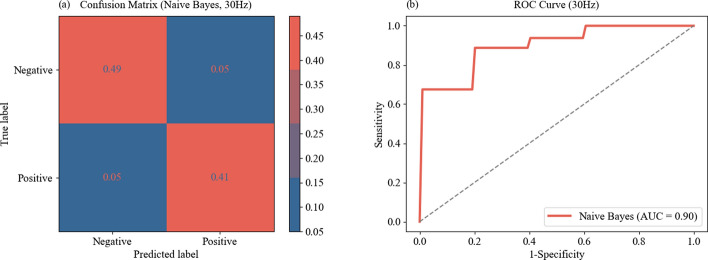
We found that BWE intensity, β band power, and γ band power exhibit significant differences across different states of alertness. The area under the receiver operating characteristic curve (AUC) of individual features for classifying alertness states was between 0.62-0.83. To further improve classification efficacy, these three features were used as input parameters in machine learning models. We found that Naive Bayes model showed the best classification efficacy in 30 Hz optical stimulation, with AUC reaching 0.90, an average accuracy of 0.90, an average sensitivity of 0.89, and an average specificity of 0.90. Meanwhile, we observed that the subjects' alertness levels did not change significantly before and after optical stimulation-induced BWE.
Our study demonstrated that the use of machine learning to integrate EEG features during 30 s optical stimulation-induced BWE showed promising classification capabilities for alertness states. It provided a rapid, quantifiable, and easily implementable alertness assessment option.
警觉性在重要任务的完成中起着至关重要的作用。然而,由于存在主观性高、练习效应、易受干扰以及数据收集复杂等问题,现有评估警觉性的方法应用受到限制。目前,迫切需要一种快速、可量化且易于实施的警觉性评估方法。
选择12种范围从4至48赫兹的光刺激频率,分别对40名受试者进行30秒的脑电波夹带(BWE)诱导。在前额极电极Fpz、Fp1和Fp2处记录脑电图(EEG)。使用卡罗林斯卡嗜睡量表、心理运动警觉性测试以及静息脑电图中的β波段功率,来评估光刺激诱导BWE前后的警觉性水平。分析了BWE期间九个EEG特征与不同警觉状态之间的相关性。接下来,采用包括支持向量机、朴素贝叶斯和逻辑回归在内的机器学习模型,对具有显著差异的EEG特征进行综合分析。
我们发现,BWE强度、β波段功率和γ波段功率在不同的警觉状态下存在显著差异。用于分类警觉状态的各个特征的受试者工作特征曲线(AUC)下面积在0.62 - 0.83之间。为了进一步提高分类效能,将这三个特征用作机器学习模型的输入参数。我们发现,朴素贝叶斯模型在30赫兹光刺激下显示出最佳的分类效能,AUC达到0.90,平均准确率为0.90,平均灵敏度为0.89,平均特异性为0.90。同时,我们观察到光刺激诱导BWE前后受试者的警觉性水平没有显著变化。
我们的研究表明,在30秒光刺激诱导BWE期间使用机器学习整合EEG特征,对警觉状态具有有前景的分类能力。它提供了一种快速、可量化且易于实施的警觉性评估选项。