Wang Lulu, Long Fang, Yan Keqing, Li Lutong, Gao Na, Xiao Zhen
Department of Obstetrics and Gynecology, First Affiliated Hospital of Dalian Medical University, Dalian, China.
People's Hospital of Nagqu, Nagqu, Tibet, China; First Affiliated Hospital of Dalian Medical University, Dalian, Liaoning, China.
Front Med (Lausanne). 2025 Jul 30;12:1603161. doi: 10.3389/fmed.2025.1603161. eCollection 2025.
This study aims to establish a simple and reproducible transvaginal mesh surgery rat model based on the modified pelvic organ prolapse rat model.
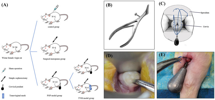
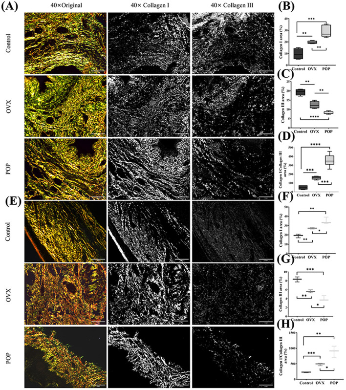
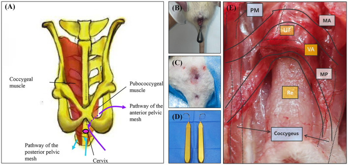
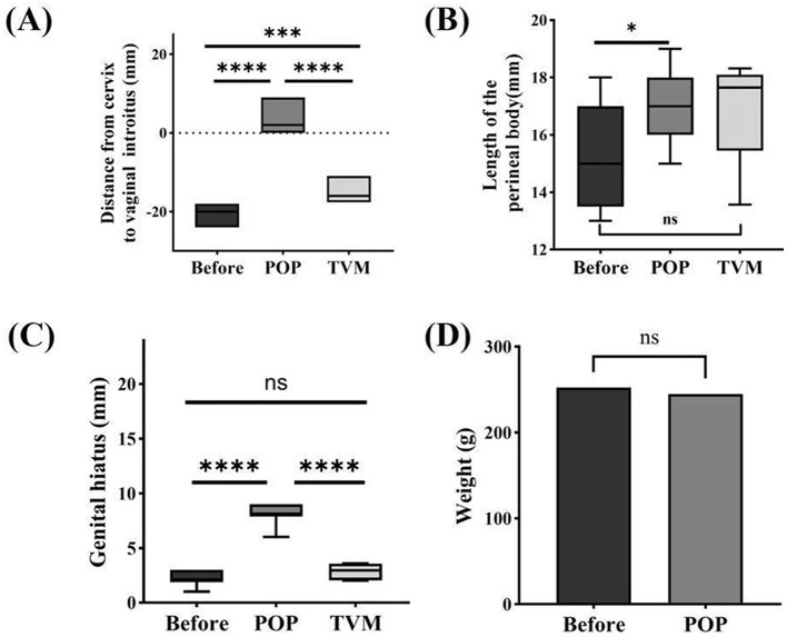
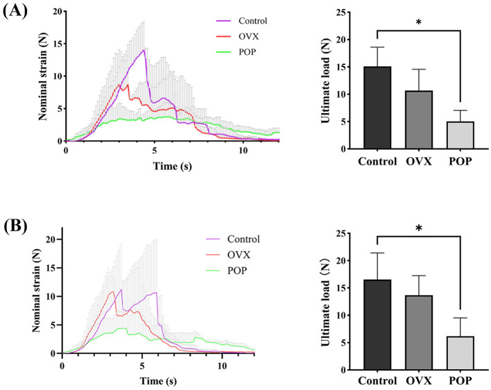
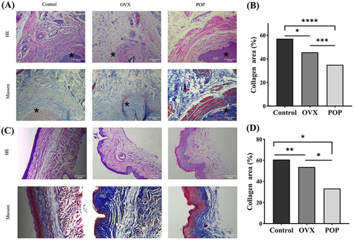
A total of 24 10-week-old female nulliparous Wistar rats were used in this study. The control group consisted of six rats with no interventions. The ovariectomy group included six rats that underwent bilateral ovariectomy. The pelvic organ prolapse group comprised 12 rats that underwent cervical pendant modeling 2 weeks after bilateral ovariectomy. Fourteen days post-modeling, six rats from the pelvic organ prolapse group underwent transvaginal mesh surgery. The rat pelvic organ prolapse quantification system was used to evaluate the prolapse condition of the rats before and after pelvic organ prolapse modeling, as well as after transvaginal mesh surgery. Vaginal wall tissue was collected to assess biomechanical changes before and after pelvic organ prolapse modeling. Additionally, vaginal wall and sacral ligament tissues were collected to evaluate structural changes and collagen alterations before and after pelvic organ prolapse modeling.
The pelvic organ prolapse rat model exhibits anatomical prolapse, biomechanical changes, and pathological changes, including collagen fiber rupture and reduced collagen density. In contrast, the transvaginal mesh rat model demonstrates anatomical recovery in prolapsed rats.
This study successfully modified the pre-existing rat model of pelvic organ prolapse and effectively mimicked human transvaginal mesh surgery using this model.
本研究旨在基于改良的盆腔器官脱垂大鼠模型建立一种简单且可重复的经阴道网片手术大鼠模型。
本研究共使用24只10周龄未生育的雌性Wistar大鼠。对照组由6只未接受干预的大鼠组成。卵巢切除组包括6只接受双侧卵巢切除的大鼠。盆腔器官脱垂组包括12只在双侧卵巢切除术后2周进行宫颈悬吊建模的大鼠。建模后14天,盆腔器官脱垂组中的6只大鼠接受经阴道网片手术。使用大鼠盆腔器官脱垂量化系统评估大鼠在盆腔器官脱垂建模前后以及经阴道网片手术后的脱垂情况。收集阴道壁组织以评估盆腔器官脱垂建模前后的生物力学变化。此外,收集阴道壁和骶韧带组织以评估盆腔器官脱垂建模前后的结构变化和胶原改变。
盆腔器官脱垂大鼠模型表现出解剖学脱垂、生物力学变化和病理变化,包括胶原纤维断裂和胶原密度降低。相比之下,经阴道网片大鼠模型显示脱垂大鼠的解剖学恢复。
本研究成功改良了现有的盆腔器官脱垂大鼠模型,并使用该模型有效模拟了人类经阴道网片手术。