Travers Gauthier, Coulomb Louise, Aouimeur Inès, He Zhiguo, Bonnet Guillaume, Ollier Edouard, Gavet Yann, Moisan Anaick, Gain Philippe, Thuret Gilles, Maurin Corantin
Laboratory Biology, Engineering and Imaging for Ophthalmology (BiiO), Faculty of Medicine, Health Innovation Campus, Jean Monnet University, Saint-Étienne, France.
Ecole Nationale Supérieure des Mines Saint-Etienne, Université de Lyon, CNRS, UMR 5307 LGF, Centre SPIN, Saint-Etienne, 42023, France.
Sci Rep. 2025 Aug 25;15(1):31301. doi: 10.1038/s41598-025-14367-4.
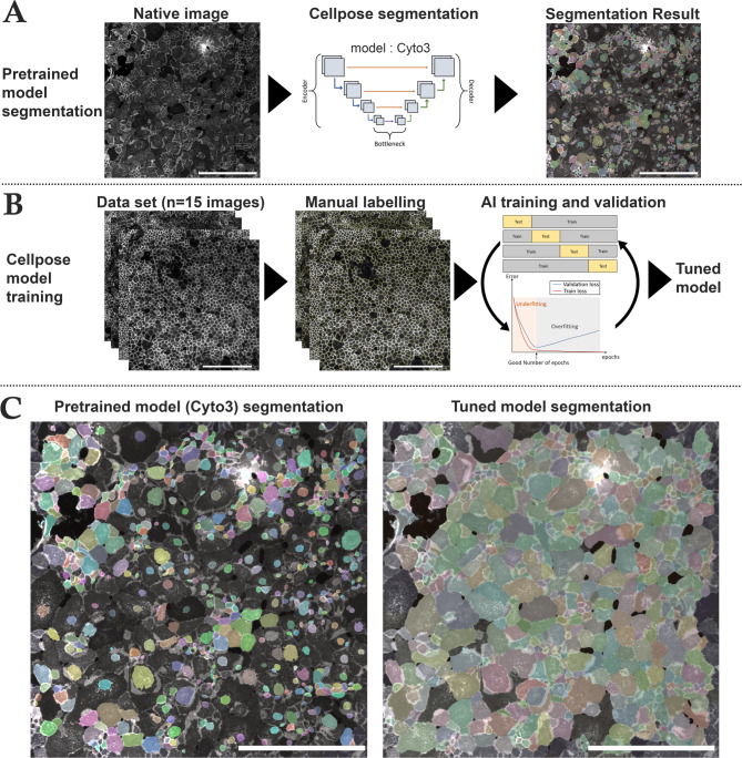
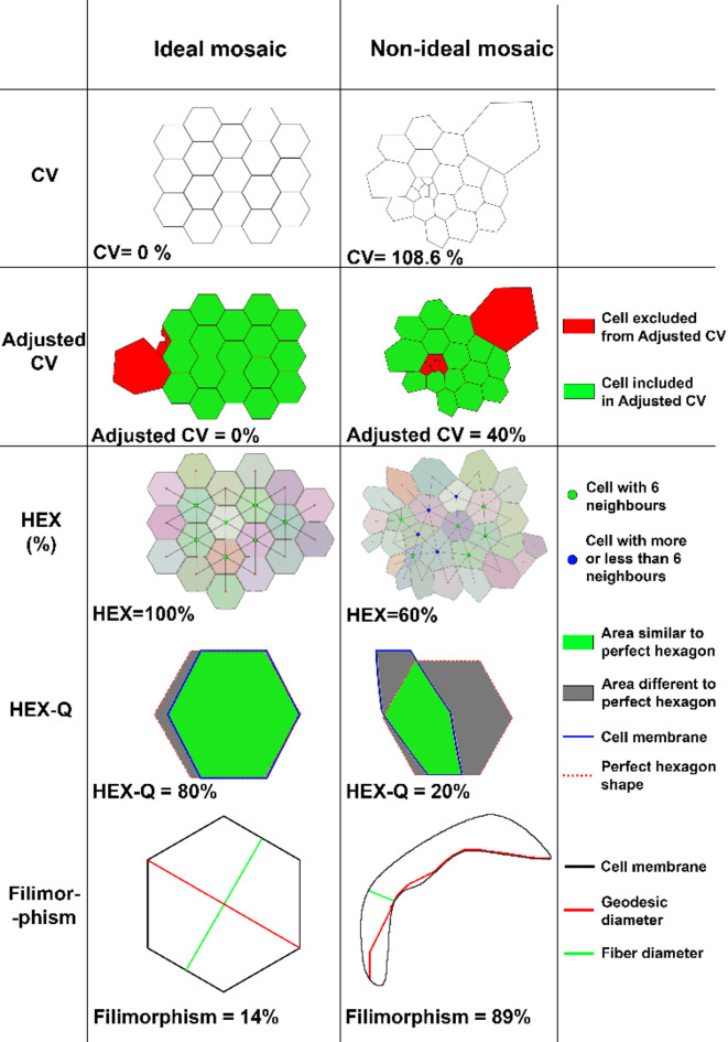
The monolayer of approximately 300,000 human corneal endothelial cells (hCECs) on the posterior surface of the cornea is essential to maintain transparency but is non-self-regenerative. Corneal blindness can currently only be treated by corneal transplantation, hindered by a global donor shortage, highlighting the need for developing tissue and/or cell therapy. The mass production of these advanced therapy medicinal products requires obtaining high-yield, high-quality endothelial cell cultures characterized by hexagonal shape, low size variability, and high endothelial cell density (ECD). Among the usual critical quality attributes which combine the expression of differentiation markers, ECD and cell morphological parameters, the latter are not optimally measured in vitro by conventional image analysis which poorly recognizes adherent cultured cells. We developed a high-performance automated segmentation using Cellpose algorithm and an original analysis method, improving the calculation of classical morphological parameters (coefficient of variation of cell area and hexagonality) and introducing new parameters specific to hCECs culture in vitro. Considering the importance of the extracellular matrix in vivo, and the panel of molecules available for coating cell culture plastics, we used these new tools to perform a comprehensive comparison of 13 molecules (laminins and collagens). We demonstrated their ability to discriminate subtle differences between cultures.
角膜后表面约300,000个人类角膜内皮细胞(hCEC)的单层对于维持透明度至关重要,但不可自我再生。目前,角膜盲只能通过角膜移植治疗,而全球供体短缺阻碍了该治疗方法,这凸显了开发组织和/或细胞疗法的必要性。这些先进治疗药物产品的大规模生产需要获得高产、高质量的内皮细胞培养物,其特征为六边形形状、低尺寸变异性和高内皮细胞密度(ECD)。在结合分化标志物表达、ECD和细胞形态学参数的常见关键质量属性中,后者在体外通过传统图像分析无法得到最佳测量,因为传统图像分析难以识别贴壁培养的细胞。我们使用Cellpose算法和一种原创分析方法开发了一种高性能自动分割方法,改进了经典形态学参数(细胞面积变异系数和六边形度)的计算,并引入了体外hCEC培养特有的新参数。考虑到细胞外基质在体内的重要性以及可用于包被细胞培养塑料的分子种类,我们使用这些新工具对13种分子(层粘连蛋白和胶原蛋白)进行了全面比较。我们证明了它们区分不同培养物之间细微差异的能力。