Rosenquist Lybecker Jenny, Van de Ven Ann, Braesch-Andersen Ken, Juriga David, Norein Norein, Hansson Per, Samanta Ayan
Macromolecular Chemistry, Department of Chemistry-Ångström Laboratory, Uppsala University, Box 538, 751 21 Uppsala, Sweden.
Thoracic Surgery, Department of Surgical Sciences, Uppsala University, 751 85 Uppsala, Sweden.
ACS Omega. 2025 Aug 14;10(33):37081-37095. doi: 10.1021/acsomega.5c01135. eCollection 2025 Aug 26.
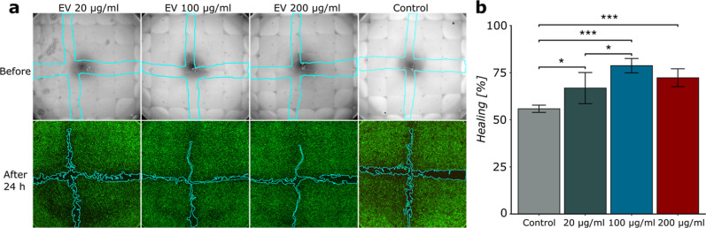
Extracellular vesicles (EVs) derived from corneal epithelial cells have shown great promise in promoting corneal wound healing and stromal regeneration, but they face challenges with rapid clearance from the eye. This study addresses these challenges by developing a biocompatible collagen-hydrogel sustained delivery system. We successfully isolated, purified, and characterized corneal epithelial EVs (CE-EVs), assessed their efficacy in corneal epithelial healing in vitro, and demonstrated their sustained delivery over 10 days followed by an on-demand release through enzymatic degradation of the hydrogel, which mimics the in vivo scenario. To develop a microscale understanding of the EV diffusion inside the hydrogel matrix, we probed the hydrogel network with several model compounds and nanoparticles by using advanced confocal microscopy analyses, followed by fitting our results to established diffusion models. Our findings suggest this innovative approach offers a safe and effective strategy to promote corneal wound healing. This technology has the potential to revolutionize corneal injury treatment and improve patient outcomes. Moreover, the possibility to tailor EV-release kinetics broadens the scope of EV research in clinical practices, as varying short- and long-term release profiles will be required for diverse medical applications.
源自角膜上皮细胞的细胞外囊泡(EVs)在促进角膜伤口愈合和基质再生方面显示出巨大潜力,但它们面临着从眼中快速清除的挑战。本研究通过开发一种生物相容性胶原水凝胶缓释系统来应对这些挑战。我们成功分离、纯化并表征了角膜上皮细胞外囊泡(CE-EVs),评估了它们在体外角膜上皮愈合中的功效,并证明它们在10天内持续释放,随后通过水凝胶的酶促降解实现按需释放,这模拟了体内情况。为了从微观层面了解细胞外囊泡在水凝胶基质中的扩散,我们使用先进的共聚焦显微镜分析,用几种模型化合物和纳米颗粒探测水凝胶网络,然后将我们的结果拟合到已建立的扩散模型中。我们的研究结果表明,这种创新方法为促进角膜伤口愈合提供了一种安全有效的策略。这项技术有可能彻底改变角膜损伤治疗方法并改善患者预后。此外,调整细胞外囊泡释放动力学的可能性拓宽了临床实践中细胞外囊泡研究的范围,因为不同的医学应用将需要不同的短期和长期释放曲线。