Sugino Noriyuki, Kitamura Yutaka, Shimada Katsumitsu, Kuroiwa Hiroko, Taguchi Akira
Department of Oral and Maxillofacial Radiology, School of Dentistry, Matsumoto Dental University, Shiojiri, JPN.
Department of Oral and Maxillofacial Surgery and Dental Implant, Center of Oral and Maxillofacial Surgery and Dental Implant in Shinshu, Obuse, JPN.
Cureus. 2025 Jul 29;17(7):e89009. doi: 10.7759/cureus.89009. eCollection 2025 Jul.
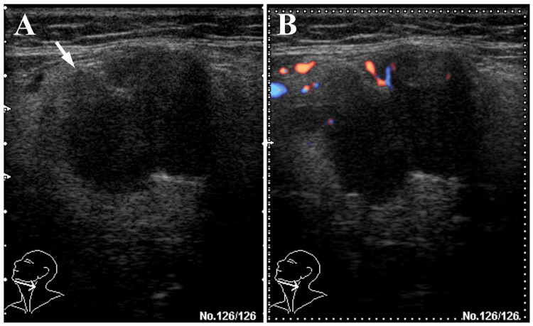
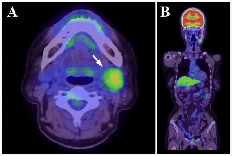
Pleomorphic adenoma (PA) is the most common benign salivary gland tumor, typically arising from the parotid gland. PA of the submandibular gland is relatively uncommon and may present diagnostic challenges, particularly when imaging findings raise suspicion of malignancy. A 66-year-old woman presented with a painless mass in the left submandibular region. Imaging studies including unenhanced computed tomography (CT), magnetic resonance imaging (MRI), ultrasonography (US), and positron emission tomography (PET) revealed a lobulated mass with irregular margins, heterogeneous internal architecture, and partially disrupted capsular structures. PET showed abnormal fluorodeoxyglucose (FDG) accumulation with a maximum standardized uptake value (SUVmax) of 3.70. Based on these findings, malignancies such as adenoid cystic carcinoma or carcinoma ex pleomorphic adenoma (CXPA) were strongly suspected. The tumor was resected under general anesthesia with careful preservation of the capsule and excised en bloc together with the submandibular gland and a portion of the sublingual gland. Histopathological and immunohistochemical analyses revealed no evidence of malignancy, and a final diagnosis of PA was made. This case highlights the diagnostic difficulty of submandibular PA, especially when capsular structures appear ambiguous on imaging. It underscores the limitations of relying solely on imaging modalities and reaffirms the importance of integrating clinical history, imaging, and pathological findings to achieve accurate diagnosis.
多形性腺瘤(PA)是最常见的涎腺良性肿瘤,通常起源于腮腺。下颌下腺的PA相对少见,可能带来诊断挑战,尤其是当影像学表现引发恶性肿瘤怀疑时。一名66岁女性患者,左侧下颌下区出现无痛性肿块。包括平扫计算机断层扫描(CT)、磁共振成像(MRI)、超声检查(US)和正电子发射断层扫描(PET)在内的影像学检查显示,肿块呈分叶状,边缘不规则,内部结构不均匀,包膜结构部分中断。PET显示异常氟脱氧葡萄糖(FDG)摄取,最大标准化摄取值(SUVmax)为3.70。基于这些发现,强烈怀疑为腺样囊性癌或多形性腺瘤恶变(CXPA)等恶性肿瘤。在全身麻醉下切除肿瘤,小心保留包膜,并与下颌下腺及部分舌下腺一并整块切除。组织病理学和免疫组化分析未发现恶性证据,最终诊断为PA。该病例凸显了下颌下腺PA的诊断困难,尤其是当包膜结构在影像学上显示不明确时。它强调了仅依靠影像学检查方法的局限性,并再次肯定了综合临床病史、影像学和病理结果以实现准确诊断的重要性。