Shen Kecheng, Sun Liang, Zhai Weiwei, Kong Lijuan, Zhu Jiandong, Yu Zhengquan, Wu Jiang
Department of Neurosurgery, The First Affiliated Hospital of Soochow University, Suzhou, Jiangsu, China.
Front Oncol. 2025 Sep 3;15:1600980. doi: 10.3389/fonc.2025.1600980. eCollection 2025.
To investigate the role of Doppler microvascular ultrasound (MVD) in skull base surgery for the intraoperative assessment and protection of arterial blood flow.
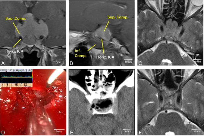
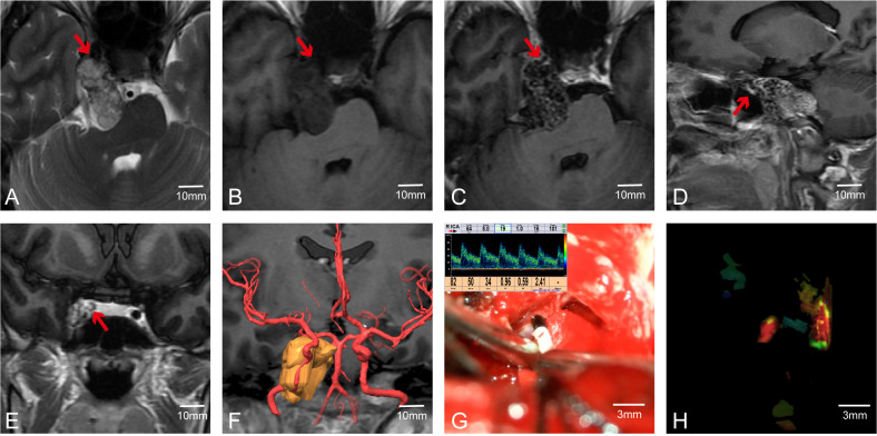
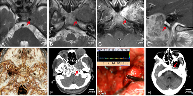
The clinical data of 56 patients who underwent surgery at the Department of Neurosurgery, First Affiliated Hospital of Soochow University between September 2017 and June 2024 were retrospectively analyzed. All patients underwent skull base tumor resection assisted by intraoperative microvascular Doppler (MVD). The procedures involved complex skull base tumors, including lesions in the sellar region, sphenoid ridge, and cavernous sinus. During surgery, MVD was utilized to monitor blood flow in vessels adjacent to the tumors. The spatial relationship and patency of the vessels and tumors were evaluated in conjunction with preoperative multimodal imaging.
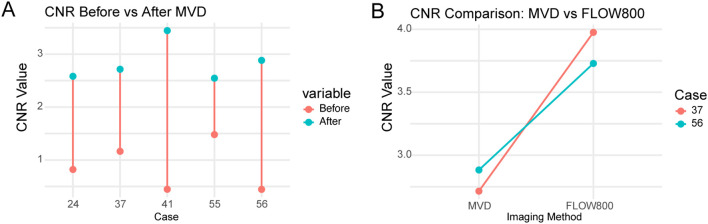
No intraoperative vascular injuries were observed among the 56 patients, confirmed by intraoperative Doppler monitoring and postoperative imaging. All patients had favorable postoperative outcomes, including no new neurological deficits. MVD facilitated precise intraoperative localization and evaluation of blood vessels, which was quantitatively supported by significant increases in contrast-to-noise ratio (CNR) after Doppler application. In two representative cases, the addition of FLOW800 fluorescence imaging provided further CNR improvement, suggesting enhanced visualization and vascular protection. Details of the CNR quantification process are provided in the Methods section.
MVD provides real-time intraoperative information on arterial blood flow during skull base surgeries, enabling surgeons to identify and preserve critical vessels, thereby improving surgical safety, resection accuracy, and patient outcomes. With ongoing technological advancements, the integration of MVD and fluorescence imaging is expected to play an increasingly vital role in complex skull base procedures.
探讨多普勒微血管超声(MVD)在颅底手术中对动脉血流进行术中评估及保护的作用。
回顾性分析2017年9月至2024年6月在苏州大学附属第一医院神经外科接受手术的56例患者的临床资料。所有患者均在术中微血管多普勒(MVD)辅助下进行颅底肿瘤切除术。手术涉及复杂的颅底肿瘤,包括鞍区、蝶骨嵴和海绵窦病变。手术过程中,利用MVD监测肿瘤邻近血管的血流情况。结合术前多模态影像评估血管与肿瘤的空间关系及通畅情况。
56例患者术中均未观察到血管损伤,术中多普勒监测及术后影像检查证实。所有患者术后预后良好,包括无新的神经功能缺损。MVD有助于术中精确地定位和评估血管,多普勒应用后对比噪声比(CNR)显著增加为其提供了定量支持。在两个典型病例中,添加FLOW800荧光成像进一步提高了CNR,表明可视化及血管保护得到增强。CNR定量过程的详细信息见方法部分。
MVD在颅底手术中提供动脉血流的术中实时信息,使外科医生能够识别并保留关键血管,从而提高手术安全性、切除准确性及患者预后。随着技术的不断进步,MVD与荧光成像的整合有望在复杂的颅底手术中发挥越来越重要的作用。