• 文献检索
  • 文档翻译
  • 深度研究
  • 学术资讯
  • Suppr Zotero 插件Zotero 插件
  • 邀请有礼
  • 套餐&价格
  • 历史记录
应用&插件
Suppr Zotero 插件Zotero 插件浏览器插件Mac 客户端Windows 客户端微信小程序
定价
高级版会员购买积分包购买API积分包
服务
文献检索文档翻译深度研究API 文档MCP 服务
关于我们
关于 Suppr公司介绍联系我们用户协议隐私条款
关注我们

Suppr 超能文献

核心技术专利:CN118964589B侵权必究
粤ICP备2023148730 号-1Suppr @ 2026

文献检索

告别复杂PubMed语法,用中文像聊天一样搜索,搜遍4000万医学文献。AI智能推荐,让科研检索更轻松。

立即免费搜索

文件翻译

保留排版,准确专业,支持PDF/Word/PPT等文件格式,支持 12+语言互译。

免费翻译文档

深度研究

AI帮你快速写综述,25分钟生成高质量综述,智能提取关键信息,辅助科研写作。

立即免费体验

新生雄性和雌性大鼠弓状核的细胞学研究

Cytology of the arcuate nucleus in newborn male and female rats.

作者信息

Walsh R J, Brawer J R

出版信息

J Anat. 1979 Jan;128(Pt 1):121-33.

PMID:422472
原文链接:https://pmc.ncbi.nlm.nih.gov/articles/PMC1232966/
Abstract

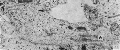
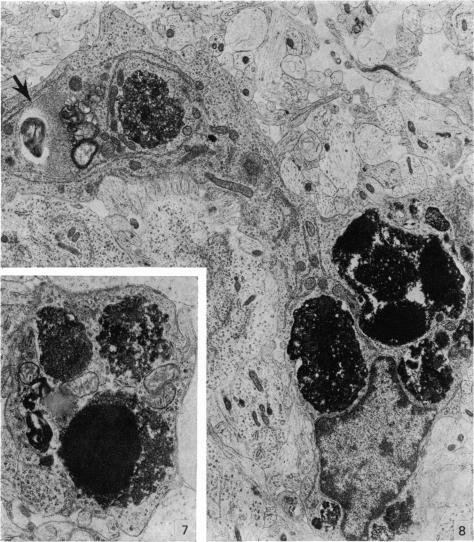
Five coronal levels of the arcuate nuclei in newborn male and female rats were examined with the transmission electron microscope. The nuclei from male and female neonates appear similar in all respects. All levels exhibit a significant population of round to oval cell profiles with large centrally located nuclei and scant cytoplasm which contains predominantly ribosomes, sparse mitochondria, and a few short cisternae of rough endoplasmic reticulum. These organelle-poor cell profiles resemble neuroblasts in other parts of the developing CNS. The arcuate nuclei of neonates also exhibit some cell profiles with the variety and quantity of organelles characteristic of mature neurons in the arcuate nuclei of adult rats. In addition, the neonatal arcuate nuclei show a paucity of synapses with apparent immaturity of those present, and numerous structures identified as growth cones. Definitive macroglia are not present in the arcuate nuclei of newborn rats.

摘要

用透射电子显微镜检查了新生雄性和雌性大鼠弓状核的五个冠状层面。雄性和雌性新生大鼠的细胞核在各方面看起来都相似。所有层面均显示出大量圆形至椭圆形的细胞轮廓,细胞核位于中央且较大,细胞质稀少,主要含有核糖体、稀疏的线粒体和一些短的粗面内质网池。这些细胞器较少的细胞轮廓类似于发育中的中枢神经系统其他部位的神经母细胞。新生大鼠的弓状核还显示出一些细胞轮廓,其细胞器的种类和数量具有成年大鼠弓状核中成熟神经元的特征。此外,新生弓状核的突触较少,且现有的突触明显不成熟,还有许多被鉴定为生长锥的结构。新生大鼠的弓状核中不存在明确的大胶质细胞。

https://cdn.ncbi.nlm.nih.gov/pmc/blobs/eac7/1232966/ae3920393ea9/janat00241-0129-c.jpg
https://cdn.ncbi.nlm.nih.gov/pmc/blobs/eac7/1232966/0b6befd77270/janat00241-0124-a.jpg
https://cdn.ncbi.nlm.nih.gov/pmc/blobs/eac7/1232966/f8e254f1960b/janat00241-0124-b.jpg
https://cdn.ncbi.nlm.nih.gov/pmc/blobs/eac7/1232966/90dc3c8d1a2c/janat00241-0124-c.jpg
https://cdn.ncbi.nlm.nih.gov/pmc/blobs/eac7/1232966/1e747d0950e0/janat00241-0126-a.jpg
https://cdn.ncbi.nlm.nih.gov/pmc/blobs/eac7/1232966/dc6cdc86738a/janat00241-0126-b.jpg
https://cdn.ncbi.nlm.nih.gov/pmc/blobs/eac7/1232966/959f600dbb5b/janat00241-0126-c.jpg
https://cdn.ncbi.nlm.nih.gov/pmc/blobs/eac7/1232966/8c94e1a58b68/janat00241-0128-a.jpg
https://cdn.ncbi.nlm.nih.gov/pmc/blobs/eac7/1232966/80f629c3ef34/janat00241-0129-a.jpg
https://cdn.ncbi.nlm.nih.gov/pmc/blobs/eac7/1232966/925120016827/janat00241-0129-b.jpg
https://cdn.ncbi.nlm.nih.gov/pmc/blobs/eac7/1232966/ae3920393ea9/janat00241-0129-c.jpg

相似文献

1
Cytology of the arcuate nucleus in newborn male and female rats.新生雄性和雌性大鼠弓状核的细胞学研究
J Anat. 1979 Jan;128(Pt 1):121-33.
2
[Comparative scanning and transmission electron microscopy studies of the ependyma of the central canal in the spinal cord of primates. I. Electron optical image of the ependyma in the central canal of the spinal cord of the callithrix monkey (Callithrix jacchus, Linné 1758)].[灵长类动物脊髓中央管室管膜的比较扫描和透射电子显微镜研究。I. 狨猴(Callithrix jacchus,林奈,1758年)脊髓中央管室管膜的电子光学图像]
Gegenbaurs Morphol Jahrb. 1986;132(4):535-54.
3
[Development of the ultrastructural organization of the supraoptic nucleus of the rat hypothalamus during postnatal ontogeny].
Arkh Anat Gistol Embriol. 1974 Sep;67(9):47-54.
4
Ultrastructural characterization of gonadotropin-releasing hormone (GnRH)-producing neurons.促性腺激素释放激素(GnRH)分泌神经元的超微结构特征
J Comp Neurol. 1985 Feb 22;232(4):534-47. doi: 10.1002/cne.902320410.
5
Histaminergic neurons in the rat brain: correlative immunocytochemistry, Golgi impregnation, and electron microscopy.大鼠脑中的组胺能神经元:相关免疫细胞化学、高尔基染色法及电子显微镜观察
J Comp Neurol. 1986 Oct 8;252(2):227-44. doi: 10.1002/cne.902520207.
6
Early postnatal development of the arcuate nucleus in normal and sexually reversed male and female rats.正常及性反转的雄性和雌性大鼠弓状核的出生后早期发育
J Anat. 1982 Dec;135(Pt 4):733-44.
7
Ultrastructural observations on the hypothalamic arcuate nuclei of aged rats in the fasting/refeeding model.禁食/再喂养模型中老年大鼠下丘脑弓状核的超微结构观察
Folia Morphol (Warsz). 2009 May;68(2):79-83.
8
Ultrastructural changes in neurons of the anterior subdivision of the rat hypothalamic ventromedial nucleus following repeated immobilization stress.
Anat Anz. 1985;158(2):125-34.
9
Ultrastructure of quiescent oocytes of Cebus albifrons.白额卷尾猴静止期卵母细胞的超微结构
J Anat. 1975 Nov;120(Pt 2):227-38.
10
Heterogeneity of the MtTw15 mammosomatotropic tumor. II. Characterization of parenchymal cells by superimposition immunocytochemistry and electron microscopy.
Anat Rec. 1980 Mar;196(3):301-11. doi: 10.1002/ar.1091960306.

引用本文的文献

1
In vivo maternal and in vitro BPA exposure effects on hypothalamic neurogenesis and appetite regulators.体内母体和体外 BPA 暴露对下丘脑神经发生和食欲调节剂的影响。
Environ Res. 2018 Jul;164:45-52. doi: 10.1016/j.envres.2018.02.011. Epub 2018 Feb 22.
2
Programmed hyperphagia secondary to increased hypothalamic SIRT1.下丘脑SIRT1增加继发的程序性摄食过量
Brain Res. 2014 Nov 17;1589:26-36. doi: 10.1016/j.brainres.2014.09.031. Epub 2014 Sep 22.
3
Fetal hypothalamic neuroprogenitor cell culture: preferential differentiation paths induced by leptin and insulin.

本文引用的文献

1
Subsurface cisterns and their relationship to the neuronal plasma membrane.脑池及其与神经元质膜的关系。
J Cell Biol. 1962 Jun;13(3):405-21. doi: 10.1083/jcb.13.3.405.
2
SEX HORMONES, BRAIN DEVELOPMENT AND BRAIN FUNCTION.性激素、大脑发育与大脑功能
Endocrinology. 1964 Oct;75:627-48. doi: 10.1210/endo-75-4-627.
3
[Electron microscopic contribution on nerve cell differentiation and histogenesis of the gray substance of the spinal cord of chick embryos].
Z Zellforsch Mikrosk Anat. 1966;74(3):401-22.
胎儿下丘脑神经前体细胞培养:瘦素和胰岛素诱导的优先分化途径。
Endocrinology. 2011 Aug;152(8):3192-201. doi: 10.1210/en.2010-1217. Epub 2011 Jun 7.
4
Hypothalamic neurosphere progenitor cells in low birth-weight rat newborns: neurotrophic effects of leptin and insulin.低出生体重新生大鼠下丘脑神经球祖细胞:瘦素和胰岛素的神经营养作用。
Brain Res. 2011 Mar 10;1378:29-42. doi: 10.1016/j.brainres.2010.12.080. Epub 2011 Jan 6.
5
Growth of fresh volumes and spontaneous cell death in the nuclei habenulae of albino rats during ontogenesis.白化大鼠个体发育过程中缰核新鲜体积的增长及自发细胞死亡
Anat Embryol (Berl). 1981;161(4):419-31. doi: 10.1007/BF00316052.
6
Early postnatal development of the arcuate nucleus in normal and sexually reversed male and female rats.正常及性反转的雄性和雌性大鼠弓状核的出生后早期发育
J Anat. 1982 Dec;135(Pt 4):733-44.
7
Postnatal development of forebrain regions in the autoimmune NZB-mouse. A model for degeneration in neuronal systems.自身免疫性NZB小鼠前脑区域的产后发育。神经元系统退化的一个模型。
Anat Embryol (Berl). 1991;183(6):579-88. doi: 10.1007/BF00187907.
4
Modification of ovulatory mechanisms by postnatal administration of estrogen to the rat.产后给大鼠注射雌激素对排卵机制的影响。
Am J Physiol. 1963 Nov;205(5):842-4. doi: 10.1152/ajplegacy.1963.205.5.842.
5
An electron microscopic study of the neuroglia during postnatal development of the rat cerebrum.大鼠大脑产后发育过程中神经胶质细胞的电子显微镜研究。
J Comp Neurol. 1968 May;133(1):45-70. doi: 10.1002/cne.901330104.
6
An electron microscopic study of neurons during postnatal development of the rat cerebral cortex.大鼠大脑皮质出生后发育过程中神经元的电子显微镜研究。
J Comp Neurol. 1968 May;133(1):17-44. doi: 10.1002/cne.901330103.
7
Studies on the developing cerebellum. Ultrastructure of the growth cones.发育中小脑的研究。生长锥的超微结构。
J Comp Neurol. 1968 Jul;133(3):341-62. doi: 10.1002/cne.901330305.
8
Developmental changes of the endoplasmic reticulum and ribosomes in nerve cells of the spinal ganglia of the domestic fowl.家鸡脊髓神经节神经细胞内质网和核糖体的发育变化
J Comp Neurol. 1968 Feb;132(2):331-64. doi: 10.1002/cne.901320207.
9
Evidence that the ovaries of the neonatal rat secrete active substances.新生大鼠卵巢分泌活性物质的证据。
J Endocrinol. 1971 Jul;50(3):529-30. doi: 10.1677/joe.0.0500529.
10
Morphology of superficial postnatal cerebral cortex with special reference to synapses.产后大脑皮层浅层的形态学,特别涉及突触
Z Zellforsch Mikrosk Anat. 1970;110(4):540-58. doi: 10.1007/BF00330103.