Goldstein J L, Wilson J D
J Clin Invest. 1972 Jul;51(7):1647-58. doi: 10.1172/JCI106966.
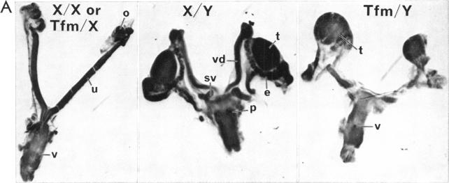
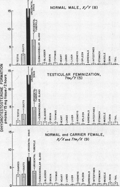
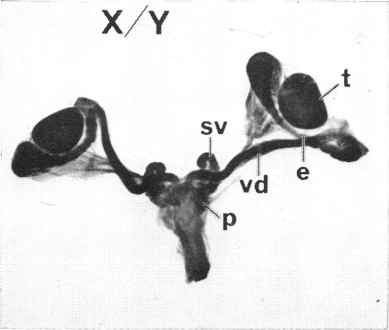
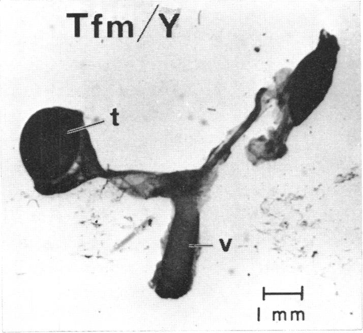
The pathogenesis of the male pseudohermaphroditism in the mouse with X-linked testicular feminization (Tfm) has been investigated by comparing testosterone formation, the effects of androgen administration, and the metabolism of testosterone-1,2-(3)H in normal mice and Tfm mice of varying ages. First, it was established that the adult Tfm animal, in contrast to the human with testicular feminization, has both a low serum testosterone and a low rate of testosterone formation as assessed in slices of testes utilizing a variety of precursors. However, the formation of testosterone from pregnenolone-7alpha-(3)H was shown to be normal in newborn Tfm testes, suggesting that a defect in testosterone synthesis may not be primary to this mutation. Second, to establish that the pseudohermaphroditic state is due to androgen resistance rather than to diminished androgen biosynthesis during fetal life, the effect of the administration of dihydrotestosterone to pregnant animals was studied in male, female, and Tfm offspring. Whereas normal and carrier female littermates demonstrated striking virilization of the internal genital tract after such treatment, there was no sign of virilization in the Tfm animals. This finding provides direct experimental evidence in support of the view that male pseudohermaphroditism in testicular feminization is the result of resistance to androgen action during androgen-mediated sexual differentiation in embryos. Third, the metabolism of testosterone-1,2-(3)H was investigated both in tissue slices and in functionally hepatectomized animals. Dihydrotestosterone formation in tissue slices of the fetal anlage of the male organs of accessory reproduction is normal in the Tfm animal, suggesting that the primary defect in this disorder involves an intracellular event subsequent to this step and that the deficient dihydrotestosterone formation observed in the adult genital tract of the Tfm mouse is secondary to the failure of differentiation in these tissues. Finally, deficient binding of testosterone in the nuclei of the submandibular gland of adult Tfm animals, a known testosterone target tissue, was demonstrated in functionally hepatectomized mice. This finding could either be a manifestation of the primary genetic defect in this disorder or might reflect another acquired abnormality due to incomplete differentiation of adrogen-sensitive cell lines.
通过比较正常小鼠和不同年龄的X连锁睾丸女性化(Tfm)小鼠的睾酮生成、雄激素给药的效果以及睾酮-1,2-(3)H的代谢,对Tfm小鼠雄性假两性畸形的发病机制进行了研究。首先,已确定成年Tfm动物与睾丸女性化的人类不同,其血清睾酮水平低,且利用多种前体在睾丸切片中评估的睾酮生成率也低。然而,在新生Tfm睾丸中,由孕烯醇酮-7α-(3)H生成睾酮显示正常,这表明睾酮合成缺陷可能不是该突变的主要原因。其次,为了确定假两性畸形状态是由于雄激素抵抗而非胎儿期雄激素生物合成减少所致,研究了给怀孕动物注射二氢睾酮对雄性、雌性和Tfm后代的影响。在此类治疗后,正常和携带基因的雌性同窝仔鼠的内生殖道显示出明显的雄性化,而Tfm动物则没有雄性化迹象。这一发现提供了直接的实验证据,支持以下观点:睾丸女性化中的雄性假两性畸形是胚胎期雄激素介导的性分化过程中对雄激素作用产生抵抗的结果。第三,在组织切片和功能性肝切除的动物中研究了睾酮-1,2-(3)H的代谢。Tfm动物辅助生殖雄性器官胎儿原基的组织切片中二氢睾酮的生成正常,这表明该疾病的主要缺陷涉及此步骤之后的细胞内事件,并且在Tfm小鼠成年生殖道中观察到的二氢睾酮生成不足是这些组织分化失败的继发结果。最后,在功能性肝切除的小鼠中证实,成年Tfm动物已知的睾酮靶组织——下颌下腺细胞核中睾酮结合不足。这一发现可能是该疾病原发性基因缺陷的表现,也可能反映了由于雄激素敏感细胞系分化不完全而导致的另一种后天异常。