• 文献检索
  • 文档翻译
  • 深度研究
  • 学术资讯
  • Suppr Zotero 插件Zotero 插件
  • 邀请有礼
  • 套餐&价格
  • 历史记录
应用&插件
Suppr Zotero 插件Zotero 插件浏览器插件Mac 客户端Windows 客户端微信小程序
定价
高级版会员购买积分包购买API积分包
服务
文献检索文档翻译深度研究API 文档MCP 服务
关于我们
关于 Suppr公司介绍联系我们用户协议隐私条款
关注我们

Suppr 超能文献

核心技术专利:CN118964589B侵权必究
粤ICP备2023148730 号-1Suppr @ 2026

文献检索

告别复杂PubMed语法,用中文像聊天一样搜索,搜遍4000万医学文献。AI智能推荐,让科研检索更轻松。

立即免费搜索

文件翻译

保留排版,准确专业,支持PDF/Word/PPT等文件格式,支持 12+语言互译。

免费翻译文档

深度研究

AI帮你快速写综述,25分钟生成高质量综述,智能提取关键信息,辅助科研写作。

立即免费体验

橙色粘球菌在粘孢子诱导过程中的结构变化。

Structural changes in Stigmatella aurantiaca during myxospore induction.

作者信息

Reichenbach H, Voelz H, Dworkin M

出版信息

J Bacteriol. 1969 Feb;97(2):905-11. doi: 10.1128/jb.97.2.905-911.1969.

DOI:10.1128/jb.97.2.905-911.1969
PMID:5773035
原文链接:https://pmc.ncbi.nlm.nih.gov/articles/PMC249776/
Abstract

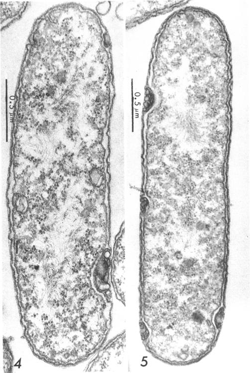
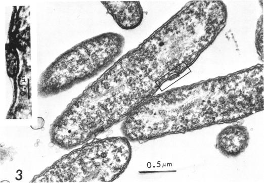
Suspension cultures of Stigmatella aurantiaca (Chondromyces aurantiacus) were induced to form myxospores by addition of glycerol to the growing culture. The cells were fixed at various stages during conversion, thin sections were prepared, and changes in fine structure were studied. Vegetative cells are quite similar in their ultrastructure to Myxococcus xanthus. During transformation into myxospores, three important cytological changes were observed. Granules of storage material, probably polysaccharide and polyphosphate, accumulated; a 200 to 300-mum thick capsule was laid down, and the outer triple layer of the cell wall became locally folded. These cell wall folds were often densely packed and lay in pockets formed by the cytoplasmic membrane. We have suggested the possibility that the cell may store in these folds wall material which has become superfluous by the decrease in surface area during conversion.

摘要

通过向生长的橙色粘球菌(软骨霉菌)悬浮培养物中添加甘油,诱导其形成粘孢子。在转化过程中的不同阶段对细胞进行固定,制备超薄切片,并研究其精细结构的变化。营养细胞的超微结构与黄色粘球菌非常相似。在转化为粘孢子的过程中,观察到三个重要的细胞学变化。储存物质颗粒,可能是多糖和多磷酸盐,积累起来;形成了一层200至300微米厚的荚膜,细胞壁的外层三层局部折叠。这些细胞壁褶皱通常紧密堆积,位于由细胞质膜形成的袋囊中。我们提出了一种可能性,即细胞可能在这些褶皱中储存由于转化过程中表面积减小而多余的细胞壁物质。

https://cdn.ncbi.nlm.nih.gov/pmc/blobs/8958/249776/13f7e156400a/jbacter00392-0466-a.jpg
https://cdn.ncbi.nlm.nih.gov/pmc/blobs/8958/249776/a4c705718d98/jbacter00392-0462-a.jpg
https://cdn.ncbi.nlm.nih.gov/pmc/blobs/8958/249776/aa8cb2fc4f0e/jbacter00392-0464-a.jpg
https://cdn.ncbi.nlm.nih.gov/pmc/blobs/8958/249776/c556a6dbc8dc/jbacter00392-0465-a.jpg
https://cdn.ncbi.nlm.nih.gov/pmc/blobs/8958/249776/13f7e156400a/jbacter00392-0466-a.jpg

相似文献

1
Structural changes in Stigmatella aurantiaca during myxospore induction.橙色粘球菌在粘孢子诱导过程中的结构变化。
J Bacteriol. 1969 Feb;97(2):905-11. doi: 10.1128/jb.97.2.905-911.1969.
2
Fine structure of fruiting bodies of Stigmatella aurantiaca (Myxobacterales).橙色粘球菌(粘杆菌目)子实体的精细结构
J Bacteriol. 1969 Sep;99(3):856-66. doi: 10.1128/jb.99.3.856-866.1969.
3
Myxospore formation in Myxococcus xanthus: chemical changes in the cell wall during cellular morphogenesis.黄色黏球菌中黏孢子的形成:细胞形态发生过程中细胞壁的化学变化
J Bacteriol. 1972 Nov;112(2):849-55. doi: 10.1128/jb.112.2.849-855.1972.
4
Induction of myxospore formation in Stigmatella aurantiaca (Myxobacterales) by monovalent cations.单价阳离子诱导橙色标桩菌(黏细菌目)形成黏孢子
J Bacteriol. 1970 Jan;101(1):325-6. doi: 10.1128/jb.101.1.325-326.1970.
5
Fruiting-body formation and myxospore differentiation and germination in Mxyococcus xanthus viewed by scanning electron microscopy.通过扫描电子显微镜观察黄色粘球菌的子实体形成、粘孢子分化及萌发过程。
J Bacteriol. 1975 Feb;121(2):711-20. doi: 10.1128/jb.121.2.711-720.1975.
6
The peptidoglycan sacculus of Myxococcus xanthus has unusual structural features and is degraded during glycerol-induced myxospore development.黄色黏球菌的肽聚糖囊具有不同寻常的结构特征,并且在甘油诱导的黏孢子发育过程中会被降解。
J Bacteriol. 2009 Jan;191(2):494-505. doi: 10.1128/JB.00608-08. Epub 2008 Nov 7.
7
Myxospore coat synthesis in Myxococcus xanthus: in vivo incorporation of acetate and glycine.黄色粘球菌中粘孢子外壳的合成:乙酸盐和甘氨酸的体内掺入
J Bacteriol. 1977 Sep;131(3):751-8. doi: 10.1128/jb.131.3.751-758.1977.
8
Acceleration of starvation- and glycerol-induced myxospore formation by prior heat shock in Myxococcus xanthus.通过先前的热休克加速饥饿和甘油诱导的黄色粘球菌形成粘孢子。
J Bacteriol. 1988 Nov;170(11):5200-7. doi: 10.1128/jb.170.11.5200-5207.1988.
9
Developmental induction of Myxococcus xanthus myxospores.黄色粘球菌粘孢子的发育诱导
J Bacteriol. 1979 Oct;140(1):147-53. doi: 10.1128/jb.140.1.147-153.1979.
10
Fine structure of Myxococcus xanthus during morphogenesis.黄色黏球菌形态发生过程中的精细结构
J Bacteriol. 1962 Nov;84(5):943-52. doi: 10.1128/jb.84.5.943-952.1962.

引用本文的文献

1
Chromosomal organization of biosynthetic gene clusters, including those of nine novel species, suggests plasticity of myxobacterial specialized metabolism.生物合成基因簇的染色体组织,包括九个新物种的基因簇,表明粘细菌特殊代谢具有可塑性。
Front Microbiol. 2023 Aug 3;14:1227206. doi: 10.3389/fmicb.2023.1227206. eCollection 2023.
2
Chromosomal organization of biosynthetic gene clusters suggests plasticity of myxobacterial specialized metabolism including descriptions for nine novel species: sp. nov., sp. nov., sp. nov., sp. nov., sp. nov., sp. nov., sp. nov., sp. nov., sp. nov.生物合成基因簇的染色体组织表明粘细菌特殊代谢具有可塑性,包括对9个新物种的描述:新物种1、新物种2、新物种3、新物种4、新物种5、新物种6、新物种7、新物种8、新物种9 。
bioRxiv. 2023 Mar 8:2023.03.08.531766. doi: 10.1101/2023.03.08.531766.
3

本文引用的文献

1
A SYSTEM FOR STUDYING MICROBIAL MORPHOGENESIS: RAPID FORMATION OF MICROCYSTS IN MYXOCOCCUS XANTHUS.一种用于研究微生物形态发生的系统:黄色粘球菌中微囊肿的快速形成。
Science. 1964 Oct 9;146(3641):243-4. doi: 10.1126/science.146.3641.243.
2
Fine structure of Myxococcus xanthus during morphogenesis.黄色黏球菌形态发生过程中的精细结构
J Bacteriol. 1962 Nov;84(5):943-52. doi: 10.1128/jb.84.5.943-952.1962.
3
The use of lead citrate at high pH as an electron-opaque stain in electron microscopy.在电子显微镜检查中,将高pH值的柠檬酸铅用作电子不透明染色剂。
Localization of the stress protein SP21 in indole-induced spores, fruiting bodies, and heat-shocked cells of Stigmatella aurantiaca.应激蛋白SP21在橙色粘球菌吲哚诱导的孢子、子实体及热激细胞中的定位
J Bacteriol. 1995 Dec;177(24):7092-9. doi: 10.1128/jb.177.24.7092-7099.1995.
4
Control of morphogenesis in Geodermatophilus: ultrastructural studies.嗜皮放线菌形态发生的控制:超微结构研究
J Bacteriol. 1970 Oct;104(1):566-80. doi: 10.1128/jb.104.1.566-580.1970.
5
Induction of myxospore formation in Stigmatella aurantiaca (Myxobacterales) by monovalent cations.单价阳离子诱导橙色标桩菌(黏细菌目)形成黏孢子
J Bacteriol. 1970 Jan;101(1):325-6. doi: 10.1128/jb.101.1.325-326.1970.
6
Fine structure of fruiting bodies of Stigmatella aurantiaca (Myxobacterales).橙色粘球菌(粘杆菌目)子实体的精细结构
J Bacteriol. 1969 Sep;99(3):856-66. doi: 10.1128/jb.99.3.856-866.1969.
7
Surface structure of gliding bacteria after freeze-etching.滑行细菌经冷冻蚀刻后的表面结构。
J Bacteriol. 1973 Jun;114(3):1351-5. doi: 10.1128/jb.114.3.1351-1355.1973.
8
Mesosomes: membranous bacterial organelles.间体:细菌的膜性细胞器。
Bacteriol Rev. 1975 Dec;39(4):405-63. doi: 10.1128/br.39.4.405-463.1975.
9
The fine structure of Herpetosiphon, and a note on the taxonomy of the genus.赫氏吸管菌的精细结构及该属分类学的一则说明。
Arch Microbiol. 1975 Mar 10;102(3):281-91. doi: 10.1007/BF00428379.
10
Ultrastructure of Polyangium cellulosum.纤维多囊菌的超微结构
J Bacteriol. 1976 Jun;126(3):1278-84. doi: 10.1128/jb.126.3.1278-1284.1976.
J Cell Biol. 1963 Apr;17(1):208-12. doi: 10.1083/jcb.17.1.208.
4
Nutritional requirements for vegetative growth of Myxococcus xanthus.黄色黏球菌营养生长的营养需求。
J Bacteriol. 1962 Aug;84(2):250-7. doi: 10.1128/jb.84.2.250-257.1962.
5
Improvements in epoxy resin embedding methods.环氧树脂包埋方法的改进。
J Biophys Biochem Cytol. 1961 Feb;9(2):409-14. doi: 10.1083/jcb.9.2.409.
6
[Electron microscopic study on plasmas containing desoxyribonucleic acid. I. Nucleoids of actively growing bacteria].[含脱氧核糖核酸血浆的电子显微镜研究。I. 活跃生长细菌的拟核]
Z Naturforsch B. 1958 Sep;13B(9):597-605.
7
The fate of the cell envelopes of Myxococcus xanthus during microcyst germination.
Arch Mikrobiol. 1966 Nov 11;55(2):110-5. doi: 10.1007/BF00418633.
8
The "polyphosphate overplus" phenomenon in Myxococcus xanthus and its influence on the architecture of the cell.黄色黏球菌中的“多聚磷酸盐过剩”现象及其对细胞结构的影响。
Arch Mikrobiol. 1966 May 9;53(4):371-88. doi: 10.1007/BF00409874.
9
The physical organization of the cytoplasm in Myxococcus zanthus and the fine structure of its components.黄色粘球菌细胞质的物理组织及其组成部分的精细结构。
Arch Mikrobiol. 1967 Jun 6;57(2):181-95. doi: 10.1007/BF00408700.
10
Fixation of ejaculated spermatozoa for electron microscopy.用于电子显微镜检查的射出精子的固定
Nature. 1967 Oct 14;216(5111):173-4. doi: 10.1038/216173a0.