Knutton S, Lloyd D R, Candy D C, McNeish A S
Infect Immun. 1984 May;44(2):519-27. doi: 10.1128/iai.44.2.519-527.1984.
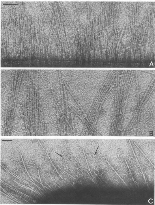
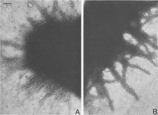
The adhesion to erythrocytes and human intestinal epithelial cells of enterotoxigenic Escherichia coli strains H10407, B2C, and H10407P, expressing colonization factor antigen I (CFA/I), CFA/II, and type 1 fimbriae, respectively, was examined by electron microscopy. CFA and type 1 fimbriae were visualized by negative staining in thin sections after en bloc staining with ruthenium red and by immune labeling with antisera raised against purified fimbriae. By negative and ruthenium red staining, CFA/I, CFA/II, and type 1 fimbriae were indistinguishable and appeared as approximately 7-nm-diameter hollow cylindrical structures up to 1.5 micron in length; strain B2C also produced 2- to 3-nm-diameter flexible fibrillar fimbriae. Bacteria producing CFA/I, CFA/II, and type 1 fimbriae adhered to and agglutinated human, bovine, and guinea pig erythrocytes, respectively; CFA/I and CFA/II also mediated attachment of bacteria to the brush border of isolated human duodenal enterocytes. Electron microscopy of agglutinated erythrocytes and enterocytes with adherent bacteria showed, in each case, that bacterial adhesion involved the formation of many interactions between the tips of fimbriae and receptors on the erythrocyte or enterocyte brush border membrane. Immune labeling allowed different fimbrial antigens mediating bacterial attachment to human enterocytes to be identified.
通过电子显微镜检查了分别表达定居因子抗原I(CFA/I)、CFA/II和1型菌毛的产肠毒素大肠杆菌菌株H10407、B2C和H10407P对红细胞和人肠上皮细胞的黏附情况。在用钌红进行整体染色后,通过负染色在薄切片中观察CFA和1型菌毛,并通过用针对纯化菌毛产生的抗血清进行免疫标记来观察。通过负染色和钌红染色,CFA/I、CFA/II和1型菌毛无法区分,呈现为直径约7纳米、长度达1.5微米的中空圆柱形结构;菌株B2C还产生直径为2至3纳米的柔性纤维状菌毛。产生CFA/I、CFA/II和1型菌毛的细菌分别与人、牛和豚鼠的红细胞黏附并凝集;CFA/I和CFA/II也介导细菌与分离的人十二指肠肠细胞刷状缘的附着。对凝集的红细胞和带有黏附细菌的肠细胞进行电子显微镜观察表明,在每种情况下,细菌黏附都涉及菌毛尖端与红细胞或肠细胞刷状缘膜上受体之间形成许多相互作用。免疫标记可鉴定介导细菌与人肠细胞附着的不同菌毛抗原。