Carlemalm E, Kellenberger E
EMBO J. 1982;1(1):63-7. doi: 10.1002/j.1460-2075.1982.tb01125.x.
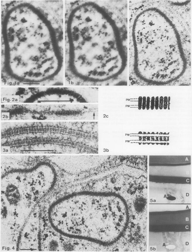
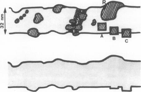
The contrast on micrographs obtained by conventional imaging in the conventional transmission electron microscope and in the scanning transmission electron microscope (STEM) (brightfield and darkfield) reflects mainly the variations of the mass-density and of the thickness of the specimen. The density differences in resin-embedded, unstained materials are too small to give enough contrast when compared to that produced by the surface perturbations introduced by sectioning. By darkfield imaging, therefore, this variable surface relief does not lead reproducibly to interpretable micrographs of high quality. Imaging by the ratio of elastically over inelastically scattered electrons in the STEM (Z-contrast) depends primarily on the atomic composition of the material. We present here the first experimental tests of theoretical predictions with thin sections; Z-contrast micrographs of septate junctions reveal the transmembrane proteins which are not visible in uranyl acetate stained sections viewed by conventional brightfield imaging.
在传统透射电子显微镜和扫描透射电子显微镜(STEM)中通过传统成像获得的显微照片上的对比度(明场和暗场)主要反映了样品质量密度和厚度的变化。与切片引入的表面扰动所产生的对比度相比,树脂包埋、未染色材料中的密度差异太小,无法提供足够的对比度。因此,通过暗场成像,这种可变的表面起伏并不能可靠地产生高质量的可解释显微照片。STEM中弹性散射电子与非弹性散射电子的比率成像(Z对比度)主要取决于材料的原子组成。我们在此展示了对薄片理论预测的首次实验测试;隔膜连接的Z对比度显微照片揭示了在传统明场成像观察的醋酸双氧铀染色切片中不可见的跨膜蛋白。