• 文献检索
  • 文档翻译
  • 深度研究
  • 学术资讯
  • Suppr Zotero 插件Zotero 插件
  • 邀请有礼
  • 套餐&价格
  • 历史记录
应用&插件
Suppr Zotero 插件Zotero 插件浏览器插件Mac 客户端Windows 客户端微信小程序
定价
高级版会员购买积分包购买API积分包
服务
文献检索文档翻译深度研究API 文档MCP 服务
关于我们
关于 Suppr公司介绍联系我们用户协议隐私条款
关注我们

Suppr 超能文献

核心技术专利:CN118964589B侵权必究
粤ICP备2023148730 号-1Suppr @ 2026

文献检索

告别复杂PubMed语法,用中文像聊天一样搜索,搜遍4000万医学文献。AI智能推荐,让科研检索更轻松。

立即免费搜索

文件翻译

保留排版,准确专业,支持PDF/Word/PPT等文件格式,支持 12+语言互译。

免费翻译文档

深度研究

AI帮你快速写综述,25分钟生成高质量综述,智能提取关键信息,辅助科研写作。

立即免费体验

传导组织的手术解剖学

The surgical anatomy of the conduction tissues.

作者信息

Anderson R H, Ho S Y, Becker A E

出版信息

Thorax. 1983 Jun;38(6):408-20. doi: 10.1136/thx.38.6.408.

DOI:10.1136/thx.38.6.408
PMID:6348996
原文链接:https://pmc.ncbi.nlm.nih.gov/articles/PMC459576/
Abstract

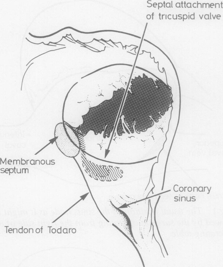
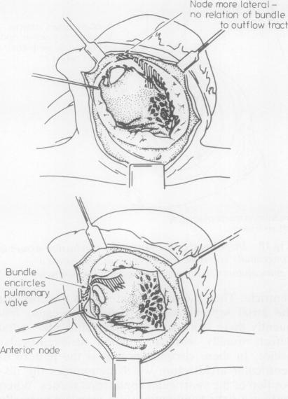
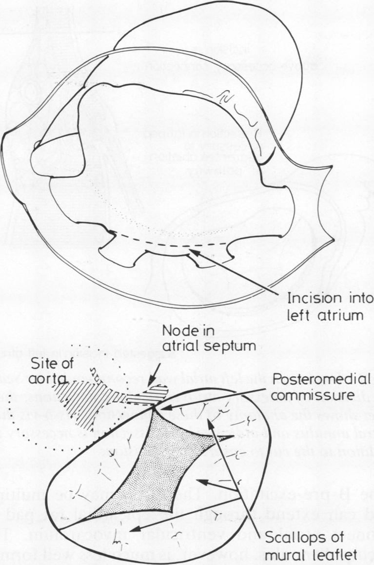
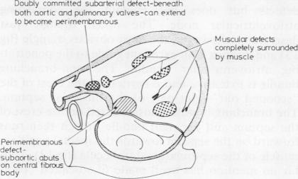
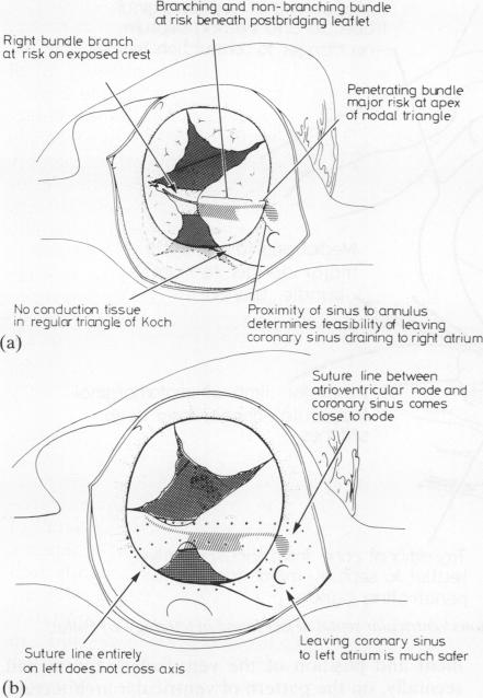
On the basis of our collective experience we have reviewed the disposition of the cardiac conduction tissues as they might be observed by the surgeon in both normal and abnormal hearts. The sinus node lies subepicardially in the terminal sulcus; because of its variable blood supply the entire superior cavoatrial junction is a potential danger area. There are no morphologically discrete tracts extending through the atrial tissues between sinus and atrioventricular nodes. The atrioventricular node, the atrial extent of the atrioventricular conduction axis, is contained exclusively within the triangle of Koch. The axis penetrates through the central fibrous body and branches on the muscular ventricular septum immediately beneath the interventricular component of the membranous septum. The landmarks to these structures are described as they might be seen through the right atrium, left atrium, and aorta. Consideration is then given to the surgical anatomy of the abnormal muscular atrioventricular connections that underscore the ventricular pre-excitation syndromes. Finally, rules are developed whereby the disposition of the conduction tissues can be predicted with accuracy in congenitally malformed hearts, in the settings of both normal and abnormal chamber connections. The most important variables in this respect are alignment between the atrial and ventricular septal structures and the pattern of ventricular architecture present.

摘要

基于我们的共同经验,我们回顾了心脏传导组织的分布情况,这些情况可由外科医生在正常和异常心脏中观察到。窦房结位于终沟的心外膜下;由于其血液供应多变,整个上腔静脉心房交界处是一个潜在的危险区域。在窦房结和房室结之间没有形态上离散的束穿过心房组织。房室结,即房室传导轴的心房部分,完全包含在科赫三角内。该轴穿过中心纤维体,并在膜性间隔的心室部分下方的肌性室间隔上分支。描述了从右心房、左心房和主动脉可能看到的这些结构的标志。然后考虑了强调心室预激综合征的异常肌性房室连接的手术解剖。最后,制定了规则,据此可以在先天性心脏畸形中,在正常和异常腔室连接的情况下准确预测传导组织的分布。在这方面最重要的变量是心房和心室间隔结构之间的对齐以及存在的心室结构模式。

https://cdn.ncbi.nlm.nih.gov/pmc/blobs/849d/459576/75c23ded6076/thorax00210-0018-a.jpg
https://cdn.ncbi.nlm.nih.gov/pmc/blobs/849d/459576/6cf71742f3a8/thorax00210-0010-a.jpg
https://cdn.ncbi.nlm.nih.gov/pmc/blobs/849d/459576/b30dfdbc02ae/thorax00210-0010-b.jpg
https://cdn.ncbi.nlm.nih.gov/pmc/blobs/849d/459576/781b674a50ca/thorax00210-0011-a.jpg
https://cdn.ncbi.nlm.nih.gov/pmc/blobs/849d/459576/a9142fde3ec6/thorax00210-0013-a.jpg
https://cdn.ncbi.nlm.nih.gov/pmc/blobs/849d/459576/daabb0b8b419/thorax00210-0013-b.jpg
https://cdn.ncbi.nlm.nih.gov/pmc/blobs/849d/459576/41f8db5729ef/thorax00210-0015-a.jpg
https://cdn.ncbi.nlm.nih.gov/pmc/blobs/849d/459576/2340d69805c1/thorax00210-0016-a.jpg
https://cdn.ncbi.nlm.nih.gov/pmc/blobs/849d/459576/bc30ea16ccc7/thorax00210-0016-b.jpg
https://cdn.ncbi.nlm.nih.gov/pmc/blobs/849d/459576/9639442a8400/thorax00210-0017-a.jpg
https://cdn.ncbi.nlm.nih.gov/pmc/blobs/849d/459576/75c23ded6076/thorax00210-0018-a.jpg

相似文献

1
The surgical anatomy of the conduction tissues.传导组织的手术解剖学
Thorax. 1983 Jun;38(6):408-20. doi: 10.1136/thx.38.6.408.
2
The architecture of the sinus node, the atrioventricular conduction axis, and the internodal atrial myocardium.窦房结、房室传导轴和结间心房肌的结构。
J Cardiovasc Electrophysiol. 1998 Nov;9(11):1233-48. doi: 10.1111/j.1540-8167.1998.tb00097.x.
3
Modification of the precise relationship of the atrioventricular conduction bundle to the margins of the ventricular septal defects by the trabecula septomarginalis.室间隔边缘小梁对房室传导束与室间隔缺损边缘精确关系的改变。
J Thorac Cardiovasc Surg. 1984 Apr;87(4):605-15.
4
The atrioventricular conduction axis of hearts with isolated ventricular septal defects.患有孤立性室间隔缺损的心脏的房室传导轴。
J Formos Med Assoc. 1990 Nov;89(11):997-1003.
5
Surgical anatomy of atrioventricular septal defect.房室间隔缺损的外科解剖学
Asian Cardiovasc Thorac Ann. 2008 Dec;16(6):497-502. doi: 10.1177/021849230801600616.
6
[Anatomy of cardiac nodes and atrioventricular specialized conduction system].[心脏节点与房室特殊传导系统的解剖结构]
Rev Esp Cardiol. 2003 Nov;56(11):1085-92. doi: 10.1016/s0300-8932(03)77019-5.
7
Atrioventricular conduction tissues in univentricular hearts of left ventricular type.左心室型单心室心脏中的房室传导组织。
Herz. 1979 Apr;4(2):166-75.
8
The AV junction region of the heart: a comprehensive study correlating gross anatomy and direct three-dimensional analysis. Part I. Architecture and topography.心脏房室交界区:一项将大体解剖与直接三维分析相关联的综合研究。第一部分。结构与局部解剖。
Anat Rec. 1999 Sep 1;256(1):49-63. doi: 10.1002/(SICI)1097-0185(19990901)256:1<49::AID-AR7>3.0.CO;2-L.
9
Anatomy of the conduction tissues 100 years on: what have we learned?百年心脏传导系统解剖学研究进展:我们学到了什么?
Heart. 2022 Aug 25;108(18):1430-1437. doi: 10.1136/heartjnl-2021-320304.
10
The conducting tissues in primitive ventricular hearts without an outlet chamber.没有流出腔的原始心室心脏中的传导组织。
Circulation. 1976 Jun;53(6):930-8. doi: 10.1161/01.cir.53.6.930.

引用本文的文献

1
Ventricular Septation of the Double-Inlet Ventricle: Over Three Decades of Follow-Up.双入口心室的室间隔分隔:三十多年的随访
Pediatr Cardiol. 2025 Apr;46(4):874-883. doi: 10.1007/s00246-024-03510-z. Epub 2024 Jun 15.
2
Intraoperative characterization of cardiac tissue: the potential of light scattering spectroscopy.术中心脏组织特征描述:光散射光谱技术的潜力。
J Biomed Opt. 2024 Jun;29(6):066005. doi: 10.1117/1.JBO.29.6.066005. Epub 2024 Jun 5.
3
3D models of the cardiac conduction system in healthy neonatal human hearts.

本文引用的文献

1
THE CONNECTING PATHWAYS BETWEEN THE SINUS NODE AND A-V NODE AND BETWEEN THE RIGHT AND THE LEFT ATRIUM IN THE HUMAN HEART.人类心脏中窦房结与房室结之间以及右心房与左心房之间的连接通路。
Am Heart J. 1963 Oct;66:498-508. doi: 10.1016/0002-8703(63)90382-x.
2
Anatomy of the atrioventricular conduction system in ventricular septal defect.室间隔缺损时房室传导系统的解剖结构。
Circulation. 1963 Jul;28:72-81. doi: 10.1161/01.cir.28.1.72.
3
Classification of ventricular septal defects.室间隔缺损的分类。
健康新生儿人心传导系统的 3D 模型。
Cardiovasc Pathol. 2024 May-Jun;70:107626. doi: 10.1016/j.carpath.2024.107626. Epub 2024 Mar 6.
4
Conduction system pacing in pediatric and congenital heart disease.小儿及先天性心脏病中的传导系统起搏
Front Physiol. 2023 Mar 24;14:1154629. doi: 10.3389/fphys.2023.1154629. eCollection 2023.
5
Editorial: Interplay between the heart and the immune system: Focus on heart rhythm regulation.社论:心脏与免疫系统之间的相互作用:关注心律调节。
Front Physiol. 2022 Aug 10;13:981499. doi: 10.3389/fphys.2022.981499. eCollection 2022.
6
Systematic Mapping of Conduction Tissue Regions During Congenital Heart Surgery.先天性心脏病手术中传导组织区域的系统映射。
Ann Thorac Surg. 2022 Oct;114(4):1500-1504. doi: 10.1016/j.athoracsur.2022.05.040. Epub 2022 Jun 11.
7
Anatomy for ablation of atrioventricular nodal reentry tachycardia and accessory pathways.房室结折返性心动过速和旁道消融的解剖学
Herzschrittmacherther Elektrophysiol. 2022 Jun;33(2):133-147. doi: 10.1007/s00399-022-00860-0. Epub 2022 May 24.
8
Back to basics: a rare and aggressive case of endocarditis.回归基础:一例罕见且侵袭性强的心内膜炎病例。
Oxf Med Case Reports. 2021 Jun 18;2021(6):omab043. doi: 10.1093/omcr/omab043. eCollection 2021 Jun.
9
Left Bundle Branch Pacing: Current Knowledge and Future Prospects.左束支起搏:当前认知与未来展望。
Front Cardiovasc Med. 2021 Mar 23;8:630399. doi: 10.3389/fcvm.2021.630399. eCollection 2021.
10
Anatomy of Mitral Valve Complex as Revealed by Non-Invasive Imaging: Pathological, Surgical and Interventional Implications.无创成像揭示的二尖瓣复合体解剖结构:病理、手术及介入意义
J Cardiovasc Dev Dis. 2020 Nov 4;7(4):49. doi: 10.3390/jcdd7040049.
Br Heart J. 1980 Mar;43(3):332-43. doi: 10.1136/hrt.43.3.332.
4
Mechanisms of cardiac arrhythmias after the Mustard operation for transposition of the great arteries.大动脉转位Mustard手术后心律失常的机制。
Am J Cardiol. 1980 Jun;45(6):1225-30. doi: 10.1016/0002-9149(80)90482-8.
5
Atrioventricular conduction system in univentricular heart of right ventricular type with right-sided rudimentary chamber.右侧有发育不全腔室的右心室型单心室心脏的房室传导系统。
Thorax. 1980 Feb;35(2):123-7. doi: 10.1136/thx.35.2.123.
6
The surgical approach to the septal area of the heart based on experiences with 45 patients with Kent bundles.基于45例肯特束患者的经验,对心脏间隔区域的手术入路。
J Thorac Cardiovasc Surg. 1980 Apr;79(4):542-51.
7
Surgical anatomy and atrioventricular conduction tissues of hearts with isolated ventricular septal defects.孤立性室间隔缺损心脏的手术解剖及房室传导组织
J Thorac Cardiovasc Surg. 1980 Feb;79(2):244-55.
8
Further observations on conduction tissues in univentricular hearts--surgical implications.单心室心脏传导组织的进一步观察——手术意义
Eur Heart J. 1981 Feb;2(1):87-96. doi: 10.1093/oxfordjournals.eurheartj.a061168.
9
Anatomico-electrophysiological correlations in the conduction system--a review.传导系统的解剖学与电生理学相关性——综述
Br Heart J. 1981 Jan;45(1):67-82. doi: 10.1136/hrt.45.1.67.
10
Surgical pathology of the conducting system of the heart.心脏传导系统的外科病理学
Br Heart J. 1967 Sep;29(5):646-70. doi: 10.1136/hrt.29.5.646.