Turner C B, Garlick P R
J Anat. 1978 Jun;126(Pt 2):385-92.
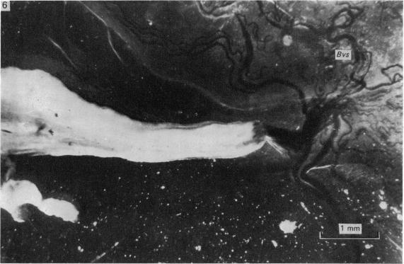
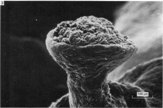
The structure of amniotic papillae in sheep was investigated by light transmission and scanning electron microscopy. Papillae were found on the amnion near the umbilical cord in a majority of the sheep examined, from early mid-term onwards. The papillae possessed a basic connective tissue core with a varying degree of vascularity, the whole being sheathed in squamous epithelial cells in the earlier stages of development; but in larger, and presumably older, papillae, squamous epithelium was absent over the tips. The blood supply to these papillae was shown to originate from the chorion and to pass into the amnion at sites where the two members were closely applied to each other. Tentatively we conclude that amniotic papillae are complex organized structures which develop near amniotic plaques, or, in some instances, from the plaques themselves. The stimuli responsible for their growth and development are unknown.
通过透光显微镜和扫描电子显微镜对绵羊羊膜乳头的结构进行了研究。从妊娠中期早期开始,在大多数被检查的绵羊中,靠近脐带的羊膜上发现了乳头。乳头具有一个基本的结缔组织核心,血管化程度各异,在发育早期整个乳头被鳞状上皮细胞包裹;但在较大且可能较老的乳头中,顶端没有鳞状上皮。这些乳头的血液供应显示源自绒毛膜,并在两层紧密贴合的部位进入羊膜。我们初步得出结论,羊膜乳头是复杂的组织结构,在羊膜斑附近发育,或者在某些情况下,由羊膜斑本身发育而来。其生长和发育的刺激因素尚不清楚。