Sutherland G R, Godman M J, Smallhorn J F, Guiterras P, Anderson R H, Hunter S
Br Heart J. 1982 Apr;47(4):316-28. doi: 10.1136/hrt.47.4.316.
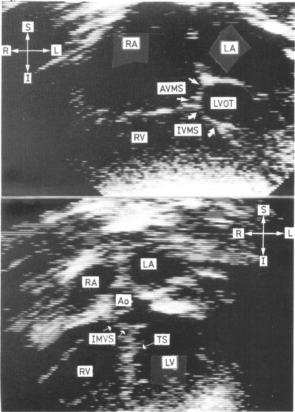
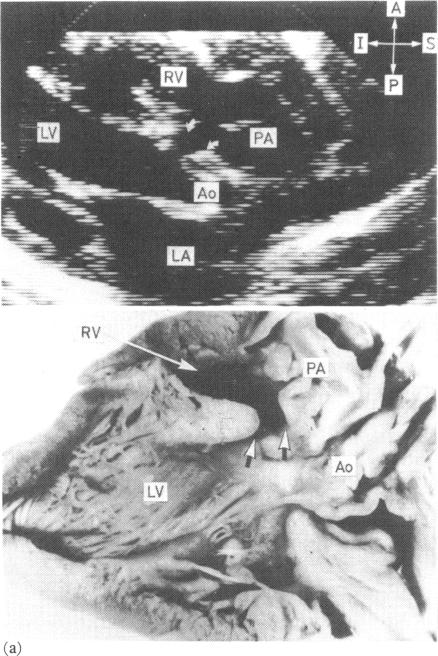
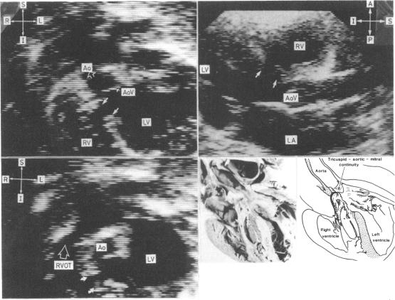
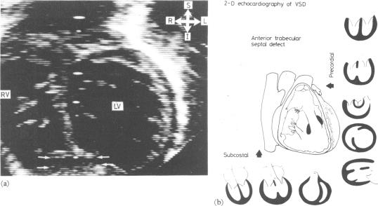
To evaluate the ability of two dimensional echocardiography to identify and classify ventricular septal defects, 280 infants and children with clinically significant ventricular septal defects were studied. Multiple precordial and subcostal echocardiographic planes were scanned in each patient in an attempt to identify the defects. Defects visualised were classified on the basis of the structures which formed their margins. Subsequent correlation of this information with angiographic (280 patients), surgical (130 patients), and pathological (31 patients) data confirmed that defects in the following sites produced a specific two dimensional echocardiographic pattern. (a) Perimembranous inlet, (b) perimembranous outlet, (c) muscular inlet, (d) single trabecular, (e) muscular outlet, and (f) doubly committed subarterial. A defect was identified and correctly classified in 252 patients. Individual defects were identified with varying degrees of accuracy. All subarterial (24 patients) defects were correctly identified and classified, as were muscular defects of the inlet (18 patients) and outlet (six patients) septa. Of the 185 perimembranous defects, 182 were identified. Only 23 of the 43 single trabecular defects were identified. Small multiple ("Swiss cheese") defects (four patients) were not identified. We conclude that two dimensional echocardiography provides a reliable non-invasive method of identifying and classifying the following ventricular septal defects: (a) perimembranous defects, (b) doubly committed subarterial defects, and (c) muscular defects of the inlet and outlet septa. In our experience it fails consistently to visualise defects in the trabecular septum.
为评估二维超声心动图识别和分类室间隔缺损的能力,对280例患有具有临床意义的室间隔缺损的婴幼儿和儿童进行了研究。对每位患者扫描多个心前区和肋下超声心动图平面,试图识别缺损。根据形成缺损边缘的结构对观察到的缺损进行分类。随后将该信息与血管造影(280例患者)、手术(130例患者)和病理(31例患者)数据进行对比,证实以下部位的缺损呈现出特定的二维超声心动图模式。(a)膜周部流入道;(b)膜周部流出道;(c)肌部流入道;(d)单小梁型;(e)肌部流出道;(f)双动脉下型。在252例患者中识别出了缺损并进行了正确分类。识别单个缺损的准确性各不相同。所有双动脉下型(24例患者)缺损以及流入道(18例患者)和流出道(6例患者)肌部缺损均被正确识别和分类。185例膜周部缺损中,识别出了182例。43例单小梁型缺损中仅识别出23例。小型多发性(“瑞士奶酪”样)缺损(4例患者)未被识别。我们得出结论,二维超声心动图提供了一种可靠的非侵入性方法来识别和分类以下室间隔缺损:(a)膜周部缺损;(b)双动脉下型缺损;(c)流入道和流出道肌部缺损。根据我们的经验,它始终无法显示小梁间隔的缺损。