Burke B, Griffiths G, Reggio H, Louvard D, Warren G
EMBO J. 1982;1(12):1621-8. doi: 10.1002/j.1460-2075.1982.tb01364.x.
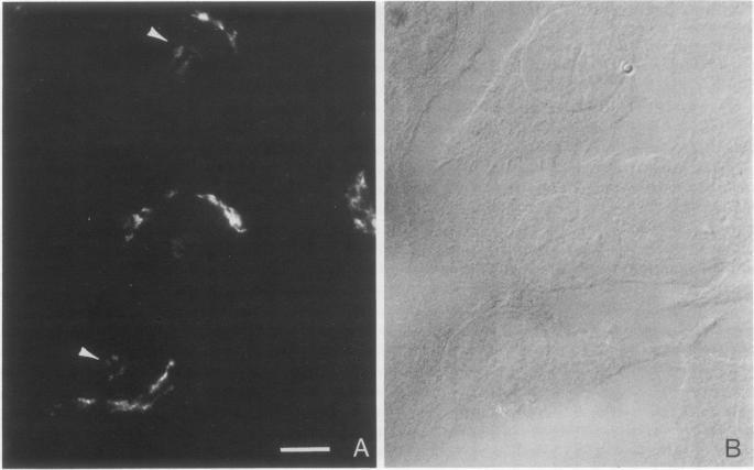
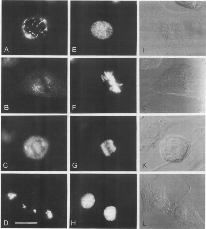
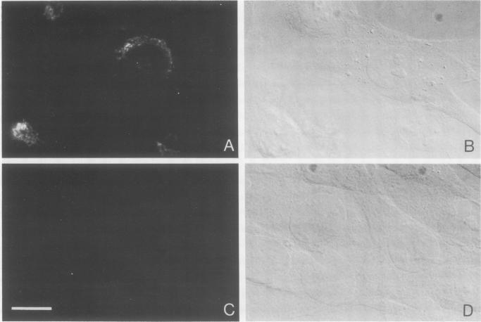
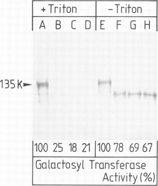
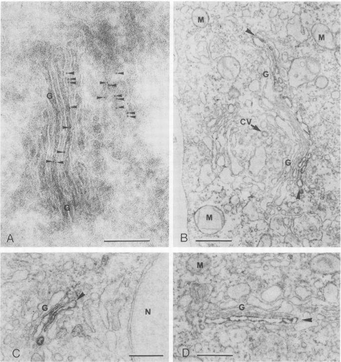
A monoclonal antibody ( 53FC3 ) has been produced against a Golgi membrane protein with a mol. wt. of 135 000 which was originally identified using a polyclonal antiserum. Treatment of isolated, intact Golgi vesicles with protease caused a decrease in mol. wt. of 5000-10 000, whereas in the presence of Triton X-100, the protein was completely degraded. This shows that the protein spans the bilayer and that most of its mass is on the luminal side of Golgi membranes. Using two immunoelectron microscopic techniques, the protein was found in one or two cisternae on one side of the Golgi stack which, in normal rat kidney cells, had 4-6 cisternae. As an illustration of the use to which this monoclonal antibody can be put we present a light microscopic study of the disassembly and reassembly of the Golgi complex during mitosis.
已制备出一种针对分子量为135000的高尔基体膜蛋白的单克隆抗体(53FC3),该蛋白最初是用多克隆抗血清鉴定出来的。用蛋白酶处理分离的完整高尔基体囊泡会导致分子量降低5000 - 10000,而在Triton X - 100存在的情况下,该蛋白会被完全降解。这表明该蛋白跨越双层膜,且其大部分质量位于高尔基体膜的腔面一侧。使用两种免疫电子显微镜技术,在正常大鼠肾细胞中由4 - 6个扁平囊组成的高尔基体堆叠一侧的一两个扁平囊中发现了该蛋白。作为这种单克隆抗体应用的一个示例,我们展示了一项关于有丝分裂期间高尔基体复合体解体和重新组装的光学显微镜研究。