Gottlieb G J, Kubo S H, Alonso D R
Am J Pathol. 1981 May;103(2):292-303.
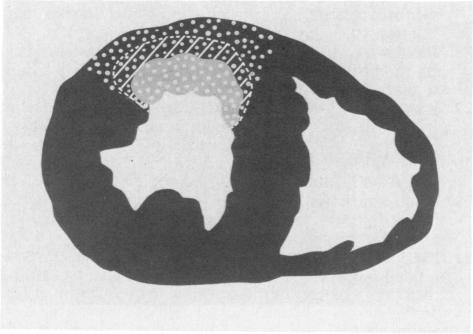
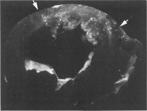
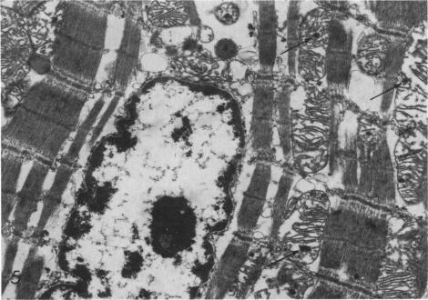
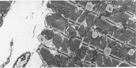
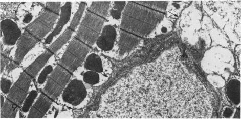
The existence of a border zone composed of reversibly injured myocardium surrounding an evolving infarct has been the subject of controversy. In experiments designed to search for such a border zone by electron microscopy, 12 mongrel dogs underwent permanent ligation of the left anterior descending coronary artery (LAD). Two to 6.5 (average = 4.2) hours later, the hearts were excised, the area at risk (myocardium perfused by the LAD) was outlined by injection of fluorescent microspheres, and the myocardial infarct was demonstrated by the nitro blue tetrazolium (NBT) gross histochemical method. Myocardial samples for electron-microscopic study were obtained from the periphery of the infarct (tissues unstained by NBT) and serially from the immediately adjacent myocardium, which was stained deep blue by NBT. Grossly, the infarcts always involved the subendocardial myocardium, extended for a variable distance in the epicardial direction, and closely approximated the lateral margins of the area at risk. When examined by electron microscopy, the infarct periphery showed evidence of irreversible damage, thus confirming the ability of NBT to detect early myocardial necrosis. Multiple samples of the NBT-stained myocardium immediately adjacent to the infarct showed varying degrees of reversible ischemia, thus demonstrating, at the ultrastructural level, the existence of a border zone of intermediate myocardial injury. This border zone was substantial (3--4 mm in width) along the subepicardial aspect of the infarct and very thin (1--2 mm) laterally. In conclusion, a significant border zone was demonstrable by electron microscopy in the subepicardial myocardium of 8 out or 12 canine hearts with recent coronary artery occlusion. In the remaining 4 hearts, the infarcts had already reached the epicardium at the time of study, and only a thin lateral border zone was present.
由围绕正在形成的梗死灶的可逆性损伤心肌组成的边界区的存在一直存在争议。在旨在通过电子显微镜寻找此类边界区的实验中,12只杂种狗接受了左冠状动脉前降支(LAD)的永久性结扎。2至6.5(平均 = 4.2)小时后,取出心脏,通过注射荧光微球勾勒出危险区域(由LAD灌注的心肌),并通过硝基蓝四氮唑(NBT)大体组织化学方法显示心肌梗死。用于电子显微镜研究的心肌样本取自梗死灶周边(未被NBT染色的组织),并依次取自紧邻的心肌,该心肌被NBT染成深蓝色。大体上,梗死灶总是累及心内膜下心肌,在心外膜方向延伸不同距离,并紧密接近危险区域的外侧边缘。通过电子显微镜检查时,梗死灶周边显示出不可逆损伤的证据,从而证实了NBT检测早期心肌坏死的能力。紧邻梗死灶的经NBT染色的心肌的多个样本显示出不同程度的可逆性缺血,从而在超微结构水平上证明了存在中间心肌损伤的边界区。该边界区在梗死灶的心外膜下方面较宽(3 - 4毫米),而在外侧非常薄(1 - 2毫米)。总之,在12只近期冠状动脉闭塞的犬心脏中,8只的心外膜下心肌通过电子显微镜可显示出明显的边界区。在其余4只心脏中,在研究时梗死灶已到达心外膜,仅存在薄的外侧边界区。