• 文献检索
  • 文档翻译
  • 深度研究
  • 学术资讯
  • Suppr Zotero 插件Zotero 插件
  • 邀请有礼
  • 套餐&价格
  • 历史记录
应用&插件
Suppr Zotero 插件Zotero 插件浏览器插件Mac 客户端Windows 客户端微信小程序
定价
高级版会员购买积分包购买API积分包
服务
文献检索文档翻译深度研究API 文档MCP 服务
关于我们
关于 Suppr公司介绍联系我们用户协议隐私条款
关注我们

Suppr 超能文献

核心技术专利:CN118964589B侵权必究
粤ICP备2023148730 号-1Suppr @ 2026

文献检索

告别复杂PubMed语法,用中文像聊天一样搜索,搜遍4000万医学文献。AI智能推荐,让科研检索更轻松。

立即免费搜索

文件翻译

保留排版,准确专业,支持PDF/Word/PPT等文件格式,支持 12+语言互译。

免费翻译文档

深度研究

AI帮你快速写综述,25分钟生成高质量综述,智能提取关键信息,辅助科研写作。

立即免费体验

成年哺乳动物骨骼肌在体外的肌生成

Myogenesis in adult mammalian skeletal muscle in vitro.

作者信息

Nag A C, Foster J D

出版信息

J Anat. 1981 Jan;132(Pt 1):1-18.

PMID:7275784
原文链接:https://pmc.ncbi.nlm.nih.gov/articles/PMC1233391/
Abstract

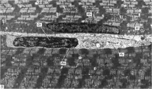
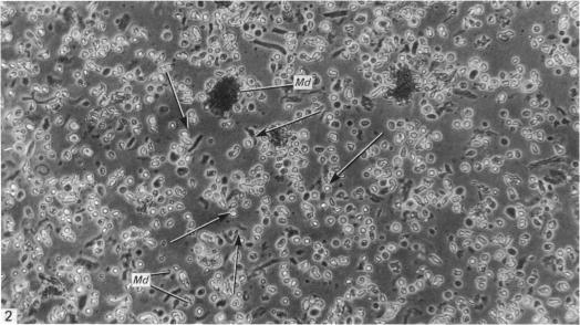
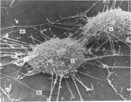
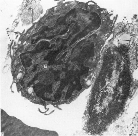
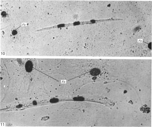
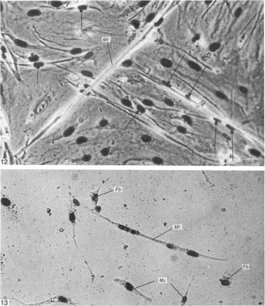
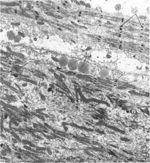
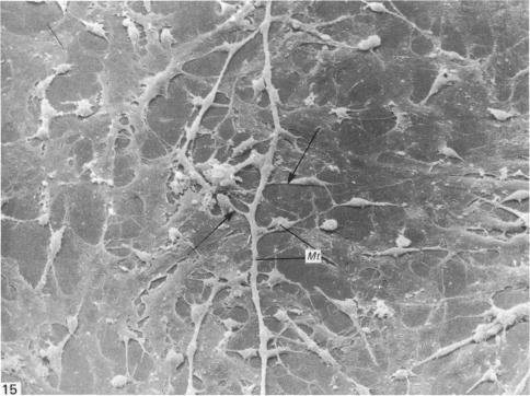
An injury to adult mammalian skeletal muscle is followed by regeneration, which involves a process believed to be similar to the differentiation of muscle fibres in the embryo. The origin of these differentiating myogenic cells is conjectural. The aim of the present study was to examine the source of myogenic cells and the process of myogenesis in adult skeletal muscle. Mononucleated cells were released from adult rat leg muscle mince after incubation with 0.1% pronase for 50-60 minutes at 37 degrees C. The ultrastructural studies revealed that the freshly dissociated mononucleated cells consisted of at least two populations of cells: myogenic satellite cells and non-myogenic fibroblastic cells. These cells were plated in growth media at various densities in cell culture dishes and incubated for 3 weeks in a balanced air atmosphere at 37 degrees C. The culture was routinely examined with a phase contrasted microscope for evidence of myogenic activities of the plated cells. At selected time intervals, the cell cultures were processed for autoradiography and scanning and transmission electron microscopy (SEM and TEM). Attachment of cells to the dish began soon after plating, with flattening of some non-muscle cells. The round- to spindle-shaped cells, indicative of myoblasts, began to appear within 24 hours. DNA synthesis and cell proliferation were observed in myogenic and non-myogenic cells within 24 hours of culture. SEM revealed that at 72 hours some myoblasts aligned and fused with one another, forming myotubes. Quantitation of autoradiographs indicated that the maximum number of labelled myotubes were present in the 3 days old culture, and thereafter, the labelled myotubes decreased in number and were absent in the 7 days old culture. Within 5-7 days the myotubes became larger and showed contractility. TEM of 6 to 21 day culture revealed that the myotubes contained well differentiated myofibrils, T-tubules and sarcoplasmic reticulum. It was evident from our studies that the mononucleated cells, having satellite cell morphology, were capable of differentiating into fully formed muscle fibres. This study lends support to the satellite cell hypothesis for regeneration of the skeletal muscle.

摘要

成年哺乳动物骨骼肌受伤后会再生,这一过程被认为与胚胎中肌纤维的分化过程相似。这些分化中的成肌细胞的起源尚无定论。本研究的目的是研究成年骨骼肌中成肌细胞的来源和肌生成过程。将成年大鼠腿部肌肉切碎,在37℃下与0.1%的链霉蛋白酶孵育50 - 60分钟后,释放出单核细胞。超微结构研究表明,刚解离的单核细胞至少由两类细胞组成:成肌卫星细胞和非成肌成纤维细胞。将这些细胞以不同密度接种于细胞培养皿中的生长培养基中,并在37℃的平衡空气环境中孵育3周。定期用相差显微镜检查培养物,以观察接种细胞的成肌活性迹象。在选定的时间间隔,对细胞培养物进行放射自显影、扫描和透射电子显微镜(SEM和TEM)处理。细胞接种后很快开始附着于培养皿,一些非肌肉细胞变扁平。圆形至梭形的细胞,即成肌细胞,在24小时内开始出现。培养24小时内,在成肌细胞和非成肌细胞中均观察到DNA合成和细胞增殖。SEM显示,72小时时一些成肌细胞相互排列并融合,形成肌管。放射自显影片的定量分析表明,标记的肌管数量在培养3天时最多,此后,标记的肌管数量减少,在培养7天时消失。在5 - 7天内,肌管变大并显示出收缩性。对培养6至21天的细胞进行TEM分析表明,肌管含有分化良好的肌原纤维、T小管和肌浆网。我们的研究表明,具有卫星细胞形态的单核细胞能够分化为完全成熟的肌纤维。本研究支持骨骼肌再生的卫星细胞假说。

https://cdn.ncbi.nlm.nih.gov/pmc/blobs/6552/1233391/b302fa96b15b/janat00225-0023-a.jpg
https://cdn.ncbi.nlm.nih.gov/pmc/blobs/6552/1233391/ed9ebd2b5a28/janat00225-0012-a.jpg
https://cdn.ncbi.nlm.nih.gov/pmc/blobs/6552/1233391/cd13f358433e/janat00225-0012-b.jpg
https://cdn.ncbi.nlm.nih.gov/pmc/blobs/6552/1233391/d43d86621286/janat00225-0013-a.jpg
https://cdn.ncbi.nlm.nih.gov/pmc/blobs/6552/1233391/cba25cafdd4e/janat00225-0014-a.jpg
https://cdn.ncbi.nlm.nih.gov/pmc/blobs/6552/1233391/ec3ad3d92a21/janat00225-0014-b.jpg
https://cdn.ncbi.nlm.nih.gov/pmc/blobs/6552/1233391/61d5743bda6b/janat00225-0016-a.jpg
https://cdn.ncbi.nlm.nih.gov/pmc/blobs/6552/1233391/5fda5b41a1af/janat00225-0017-a.jpg
https://cdn.ncbi.nlm.nih.gov/pmc/blobs/6552/1233391/46126227c829/janat00225-0017-b.jpg
https://cdn.ncbi.nlm.nih.gov/pmc/blobs/6552/1233391/29eeb8513aee/janat00225-0018-a.jpg
https://cdn.ncbi.nlm.nih.gov/pmc/blobs/6552/1233391/b489e4bc18e8/janat00225-0019-a.jpg
https://cdn.ncbi.nlm.nih.gov/pmc/blobs/6552/1233391/dcc4e150adce/janat00225-0020-a.jpg
https://cdn.ncbi.nlm.nih.gov/pmc/blobs/6552/1233391/6c241a203721/janat00225-0021-a.jpg
https://cdn.ncbi.nlm.nih.gov/pmc/blobs/6552/1233391/f4f4b8dd3d9a/janat00225-0021-b.jpg
https://cdn.ncbi.nlm.nih.gov/pmc/blobs/6552/1233391/2f5c517e4b91/janat00225-0022-a.jpg
https://cdn.ncbi.nlm.nih.gov/pmc/blobs/6552/1233391/b302fa96b15b/janat00225-0023-a.jpg

相似文献

1
Myogenesis in adult mammalian skeletal muscle in vitro.成年哺乳动物骨骼肌在体外的肌生成
J Anat. 1981 Jan;132(Pt 1):1-18.
2
Regeneration of adult newt skeletal muscle tissue in vitro.成年蝾螈骨骼肌组织的体外再生
J Embryol Exp Morphol. 1983 Oct;77:255-71.
3
The role of myonuclei in muscle regeneration: an in vitro study.肌细胞核在肌肉再生中的作用:一项体外研究。
J Cell Physiol. 1978 Aug;96(2):245-51. doi: 10.1002/jcp.1040960213.
4
Satellite cell proliferation and the expression of myogenin and desmin in regenerating skeletal muscle: evidence for two different populations of satellite cells.卫星细胞增殖以及生肌调节因子和结蛋白在再生骨骼肌中的表达:卫星细胞存在两种不同群体的证据
Lab Invest. 1995 Mar;72(3):341-7.
5
Myogenic cell formation in regenerating rat skeletal muscle injured by mincing. I. A fine structural study.
Anat Rec. 1977 Jun;188(2):181-99. doi: 10.1002/ar.1091880205.
6
Characterization of myogenesis from adult satellite cells cultured in vitro.
Int J Dev Biol. 1990 Mar;34(1):171-80.
7
Desmin is present in proliferating rat muscle satellite cells but not in bovine muscle satellite cells.结蛋白存在于增殖的大鼠肌肉卫星细胞中,但不存在于牛肌肉卫星细胞中。
J Cell Physiol. 1991 Dec;149(3):525-35. doi: 10.1002/jcp.1041490323.
8
Abortive myogenesis in denervated skeletal muscle: differentiative properties of satellite cells, their migration, and block of terminal differentiation.失神经支配骨骼肌中的顿挫性肌生成:卫星细胞的分化特性、迁移及终末分化阻滞
Anat Embryol (Berl). 2005 Apr;209(4):269-79. doi: 10.1007/s00429-004-0429-7. Epub 2005 Mar 11.
9
Myogenic cell formation in regenerating rat skeletal muscle injured by mincing. II. An autoradiographic study.
Anat Rec. 1977 Jun;188(2):201-17. doi: 10.1002/ar.1091880206.
10
[Ultrastructure of the cells and DNA synthesis in skeletal muscle regeneration. A study of the regeneration of the frog sartorius muscle by an electron microscopic autoradiographic method].[骨骼肌再生中的细胞超微结构与DNA合成。用电子显微镜放射自显影法对青蛙缝匠肌再生的研究]
Tsitologiia. 1977;19(12):1333-9.

引用本文的文献

1
3D Bioprinting and Differentiation of Primary Skeletal Muscle Progenitor Cells.3D 生物打印与原代骨骼肌祖细胞的分化。
Methods Mol Biol. 2020;2140:229-242. doi: 10.1007/978-1-0716-0520-2_15.
2
Long-Term High-Density Extracellular Recordings Enable Studies of Muscle Cell Physiology.长期高密度细胞外记录有助于肌肉细胞生理学研究。
Front Physiol. 2018 Oct 9;9:1424. doi: 10.3389/fphys.2018.01424. eCollection 2018.
3
On-chip, multisite extracellular and intracellular recordings from primary cultured skeletal myotubes.芯片上原代培养骨骼肌肌管的多点细胞外和细胞内记录。

本文引用的文献

1
Satellite cell of skeletal muscle fibers.骨骼肌纤维的卫星细胞。
J Biophys Biochem Cytol. 1961 Feb;9(2):493-5. doi: 10.1083/jcb.9.2.493.
2
Electron microscopic observations of satellite cells with special reference to the development of mammalian skeletal muscles.卫星细胞的电子显微镜观察,特别涉及哺乳动物骨骼肌的发育
Z Anat Entwicklungsgesch. 1966;125(1):43-63. doi: 10.1007/BF00521974.
3
The histogenesis of rat intercostal muscle.大鼠肋间肌的组织发生
Sci Rep. 2016 Nov 4;6:36498. doi: 10.1038/srep36498.
4
Identification and Characterization of the Dermal Panniculus Carnosus Muscle Stem Cells.真皮肉膜肌干细胞的鉴定与特征分析
Stem Cell Reports. 2016 Sep 13;7(3):411-424. doi: 10.1016/j.stemcr.2016.08.002. Epub 2016 Sep 1.
5
Muscle fibers from senescent mice retain excitation-contraction coupling properties in culture.衰老小鼠的肌纤维在培养中保留兴奋-收缩偶联特性。
In Vitro Cell Dev Biol Anim. 2007 Jul-Aug;43(7):222-34. doi: 10.1007/s11626-007-9047-z. Epub 2007 Aug 22.
6
Adult fast myosin pattern and Ca2+-induced slow myosin pattern in primary skeletal muscle culture.原代骨骼肌培养中的成人快速肌球蛋白模式和钙诱导的慢速肌球蛋白模式。
Proc Natl Acad Sci U S A. 1997 Apr 15;94(8):4205-10. doi: 10.1073/pnas.94.8.4205.
7
Ultrastructural study of developing rabbit diaphragm.发育中兔膈肌的超微结构研究
J Anat. 1984 Aug;139 ( Pt 1)(Pt 1):67-79.
8
Proliferation of myogenic cells in the diaphragm of the fetal rabbit.胎兔膈肌中肌源性细胞的增殖
Can J Vet Res. 1986 Jan;50(1):74-7.
J Cell Biol. 1969 Jul;42(1):135-53. doi: 10.1083/jcb.42.1.135.
4
Origin of myoblasts during skeletal muscle regeneration. Electron microscopic observations.
Lab Invest. 1969 Apr;20(4):353-63.
5
The satellite cell bud and myoblast in denervated mammalian muscle fibers.
Am J Anat. 1970 Sep;129(1):21-39. doi: 10.1002/aja.1001290103.
6
Satellite cells as the source of nuclei in muscles of growing rats.卫星细胞作为生长中大鼠肌肉细胞核的来源。
Anat Rec. 1971 Aug;170(4):421-35. doi: 10.1002/ar.1091700405.
7
Changes in surface morphology of Chinese hamster ovary cells during the cell cycle.中国仓鼠卵巢细胞在细胞周期中的表面形态变化。
J Cell Biol. 1973 Jun;57(3):815-36. doi: 10.1083/jcb.57.3.815.
8
Ligand-mediated osmium binding: its application in coating biological specimens for scanning electron microscopy.配体介导的锇结合:其在扫描电子显微镜生物样本包被中的应用。
J Ultrastruct Res. 1973 Nov;45(3):254-8. doi: 10.1016/s0022-5320(73)80051-6.
9
Enzymatic liberation of myogenic cells from adult rat muscle.从成年大鼠肌肉中酶促释放生肌细胞。
Anat Rec. 1974 Dec;180(4):645-61. doi: 10.1002/ar.1091800410.
10
The regeneration of skeletal muscle. A review.骨骼肌的再生。综述。
Am J Anat. 1973 Jun;137(2):119-49. doi: 10.1002/aja.1001370202.