• 文献检索
  • 文档翻译
  • 深度研究
  • 学术资讯
  • Suppr Zotero 插件Zotero 插件
  • 邀请有礼
  • 套餐&价格
  • 历史记录
应用&插件
Suppr Zotero 插件Zotero 插件浏览器插件Mac 客户端Windows 客户端微信小程序
定价
高级版会员购买积分包购买API积分包
服务
文献检索文档翻译深度研究API 文档MCP 服务
关于我们
关于 Suppr公司介绍联系我们用户协议隐私条款
关注我们

Suppr 超能文献

核心技术专利:CN118964589B侵权必究
粤ICP备2023148730 号-1Suppr @ 2026

文献检索

告别复杂PubMed语法,用中文像聊天一样搜索,搜遍4000万医学文献。AI智能推荐,让科研检索更轻松。

立即免费搜索

文件翻译

保留排版,准确专业,支持PDF/Word/PPT等文件格式,支持 12+语言互译。

免费翻译文档

深度研究

AI帮你快速写综述,25分钟生成高质量综述,智能提取关键信息,辅助科研写作。

立即免费体验

噬菌体T7和T7衣壳在琼脂糖凝胶中的电泳

Electrophoresis of bacteriophage T7 and T7 capsids in agarose gels.

作者信息

Serwer P, Pichler M E

出版信息

J Virol. 1978 Dec;28(3):917-28. doi: 10.1128/JVI.28.3.917-928.1978.

DOI:10.1128/JVI.28.3.917-928.1978
PMID:731798
原文链接:https://pmc.ncbi.nlm.nih.gov/articles/PMC525816/
Abstract

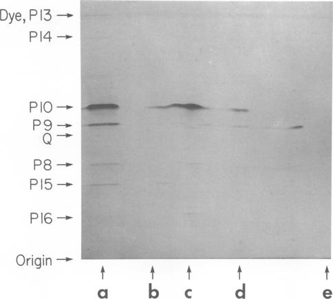
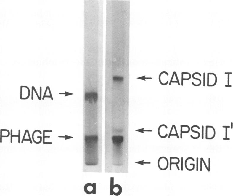
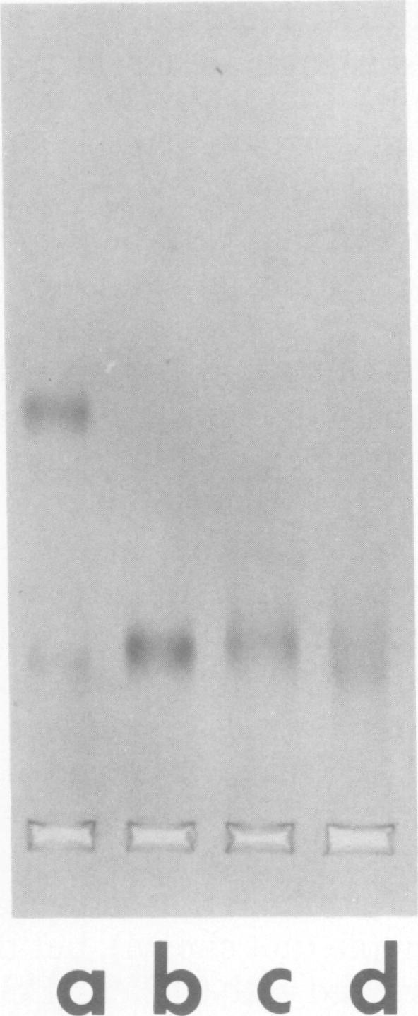
Agarose gel electrophoresis of the following was performed in 0.05 M sodium phosphate-0.001 M MgCl2 (pH 7.4): (i) bacteriophage T7; (ii) a T7 precursor capsid (capsid I), isolated from T7-infected Escherichia coli, which has a thicker and less angular envelope than bacteriophage T7; (iii) a second capsid (capsid II), isolated from T7-infected E. coli, which has a bacteriophage-like envelope; and (iv) capsids (capsid IV) produced by temperature shock of bacteriophage T7. Bacteriophage T7 and all of the above capsids migrated towards the anode. In a 0.9% agarose gel, capsid I had an electrophoretic mobility of 9.1 +/- 0.4 X 10(-5) cm2/V.s; bacteriophage T7 migrated 0.31 +/- 0.02 times as fast as capsid I. The mobilities of different preparations of capsid II varied in such gels: the fastest-migrating capsid II preparation was 0.51 +/- 0.03 times as fast as capsid I and the slowest was 0.37 +/- 0.02 times as fast as capsid I. Capsid IV with and without the phage tail migrated 0.29 +/- 0.02 and 0.42 +/- 0.02 times as fast as capsid I. The results of the extrapolation of bacteriophage and capsid mobilities to 0% agarose concentration indicated that the above differences in mobility are caused by differences in average surface charge density. To increase the accuracy of mobility comparisons and to increase the number of samples that could be simultaneously analyzed, multisample horizontal slab gels were used. Treatment with the ionic detergent sodium dodecyl sulfate converted capsid I to a capsid that migated in the capsid II region during electrophoresis through agarose gels. In the electron microscope, most of the envelopes of these latter capsids resembled the capsid II envelope, but some envelope regions were thicker than the capsid II envelope.

摘要

在0.05M磷酸钠 - 0.001M氯化镁(pH 7.4)中对以下物质进行琼脂糖凝胶电泳:(i)噬菌体T7;(ii)从T7感染的大肠杆菌中分离出的T7前衣壳(衣壳I),其包膜比噬菌体T7更厚且棱角更少;(iii)从T7感染的大肠杆菌中分离出的第二种衣壳(衣壳II),其具有类似噬菌体的包膜;以及(iv)通过噬菌体T7温度休克产生的衣壳(衣壳IV)。噬菌体T7和上述所有衣壳都向阳极迁移。在0.9%琼脂糖凝胶中,衣壳I的电泳迁移率为9.1±0.4×10⁻⁵cm²/V·s;噬菌体T7的迁移速度是衣壳I的0.31±0.02倍。不同制备的衣壳II在这种凝胶中的迁移率有所不同:迁移最快的衣壳II制剂的速度是衣壳I的0.51±0.03倍,最慢的是衣壳I的0.37±0.02倍。带有和不带有噬菌体尾部的衣壳IV的迁移速度分别是衣壳I的0.29±0.02倍和0.42±0.02倍。将噬菌体和衣壳迁移率外推至0%琼脂糖浓度的结果表明,上述迁移率差异是由平均表面电荷密度差异引起的。为了提高迁移率比较的准确性并增加可同时分析的样品数量,使用了多样品水平平板凝胶。用离子去污剂十二烷基硫酸钠处理后,衣壳I在通过琼脂糖凝胶电泳时转变为在衣壳II区域迁移的衣壳。在电子显微镜下,这些后一种衣壳的大多数包膜类似于衣壳II包膜,但一些包膜区域比衣壳II包膜更厚。

https://cdn.ncbi.nlm.nih.gov/pmc/blobs/42c5/525816/d58e2bc97bef/jvirol00204-0269-a.jpg
https://cdn.ncbi.nlm.nih.gov/pmc/blobs/42c5/525816/f02d92958e9d/jvirol00204-0262-a.jpg
https://cdn.ncbi.nlm.nih.gov/pmc/blobs/42c5/525816/7391af53f97e/jvirol00204-0263-a.jpg
https://cdn.ncbi.nlm.nih.gov/pmc/blobs/42c5/525816/d965b3a3232f/jvirol00204-0265-a.jpg
https://cdn.ncbi.nlm.nih.gov/pmc/blobs/42c5/525816/c399bc06fec3/jvirol00204-0266-a.jpg
https://cdn.ncbi.nlm.nih.gov/pmc/blobs/42c5/525816/b860d89925cd/jvirol00204-0268-a.jpg
https://cdn.ncbi.nlm.nih.gov/pmc/blobs/42c5/525816/d58e2bc97bef/jvirol00204-0269-a.jpg

相似文献

1
Electrophoresis of bacteriophage T7 and T7 capsids in agarose gels.噬菌体T7和T7衣壳在琼脂糖凝胶中的电泳
J Virol. 1978 Dec;28(3):917-28. doi: 10.1128/JVI.28.3.917-928.1978.
2
Comparison of the physical properties and assembly pathways of the related bacteriophages T7, T3 and phi II.相关噬菌体T7、T3和φII的物理性质及组装途径比较
J Mol Biol. 1983 Oct 25;170(2):447-69. doi: 10.1016/s0022-2836(83)80157-0.
3
Assembly of bacteriophage T7. Dimensions of the bacteriophage and its capsids.噬菌体T7的组装。噬菌体及其衣壳的尺寸。
Biophys J. 1981 Dec;36(3):743-57. doi: 10.1016/S0006-3495(81)84763-7.
4
Detection and characterization of agarose-binding, capsid-like particles produced during assembly of a bacteriophage T7 procapsid.噬菌体T7原衣壳组装过程中产生的琼脂糖结合衣壳样颗粒的检测与表征
J Virol. 1982 May;42(2):583-94. doi: 10.1128/JVI.42.2.583-594.1982.
5
Changes in the capsid structure and stability of defective particles of bacteriophage R17.噬菌体R17缺陷颗粒的衣壳结构和稳定性变化
J Virol. 1977 Nov;24(2):590-601. doi: 10.1128/JVI.24.2.590-601.1977.
6
A small (58-nm) attached sphere perturbs the sieving of 40-80-kilobase DNA in 0.2-2.5% agarose gels: analysis of bacteriophage T7 capsid-DNA complexes by use of pulsed field electrophoresis.一个小的(58纳米)附着球体干扰了0.2 - 2.5%琼脂糖凝胶中40 - 80千碱基DNA的筛分:利用脉冲场电泳分析噬菌体T7衣壳 - DNA复合物。
Biochemistry. 1992 Sep 15;31(36):8397-405. doi: 10.1021/bi00151a002.
7
Gel electrophoretic analysis of bacteriophage assembly intermediates in bacteriophage plaques.噬菌体噬菌斑中噬菌体组装中间体的凝胶电泳分析。
Appl Theor Electrophor. 1995;4(4):211-7.
8
Capsid-DNA complexes in the DNA packaging pathway of bacteriophage T7: characterization of the capsids bound to monomeric and concatemeric DNA.噬菌体T7 DNA包装途径中的衣壳-DNA复合物:与单体和串联体DNA结合的衣壳的特性
Virology. 1981 Jan 15;108(1):164-76. doi: 10.1016/0042-6822(81)90536-5.
9
The conformation of packaged bacteriophage T7 DNA: informative images of negatively stained T7.包装的噬菌体T7 DNA的构象:经负染色的T7的信息性图像。
J Struct Biol. 1997 Oct;120(1):32-43. doi: 10.1006/jsbi.1997.3895.
10
The identification of late bacteriophage T4 proteins on sodium dodecyl sulfate polyacrylamide gels.在十二烷基硫酸钠聚丙烯酰胺凝胶上对晚期噬菌体T4蛋白的鉴定。
Virology. 1974 Jul;60(1):265-75. doi: 10.1016/0042-6822(74)90384-5.

引用本文的文献

1
Bacteriophages in Natural and Artificial Environments.自然与人工环境中的噬菌体
Pathogens. 2019 Jul 12;8(3):100. doi: 10.3390/pathogens8030100.
2
Nanomedicine and Phage Capsids.纳米医学与噬菌体衣壳
Viruses. 2018 Jun 6;10(6):307. doi: 10.3390/v10060307.
3
Semi-Solid and Solid Dosage Forms for the Delivery of Phage Therapy to Epithelia.用于向上皮组织递送噬菌体疗法的半固体和固体剂型。

本文引用的文献

1
Molecular weights of coliphages and coliphage DNA. I. Measurement of the molecular weight of bacteriophage T7 by high-speed equilibrium centrifugation.大肠杆菌噬菌体和大肠杆菌噬菌体DNA的分子量。I. 用高速平衡离心法测定噬菌体T7的分子量。
J Mol Biol. 1970 Dec 28;54(3):537-46. doi: 10.1016/0022-2836(70)90124-5.
2
Bacteriophage T7.噬菌体T7
Science. 1972 Apr 28;176(4033):367-76. doi: 10.1126/science.176.4033.367.
3
Fast sedimenting bacteriophage T7 DNA from T7-infected Escherichia coli.来自T7感染的大肠杆菌的快速沉降噬菌体T7 DNA。
Pharmaceuticals (Basel). 2018 Feb 26;11(1):26. doi: 10.3390/ph11010026.
4
Phage-mediated counting by the naked eye of miRNA molecules at attomolar concentrations in a Petri dish.噬菌体介导的在培养皿中对阿托摩尔浓度的微小RNA分子进行肉眼计数。
Nat Mater. 2015 Oct;14(10):1058-64. doi: 10.1038/nmat4377. Epub 2015 Aug 17.
5
Capture and detection of T7 bacteriophages on a nanostructured interface.
ACS Appl Mater Interfaces. 2014 Apr 9;6(7):4758-65. doi: 10.1021/am500655r. Epub 2014 Mar 28.
6
Viral information.病毒信息。
Biol Philos. 2013 Mar;28(2):283-297. doi: 10.1007/s10539-012-9344-0. Epub 2012 Oct 31.
7
Structural evolution of the P22-like phages: comparison of Sf6 and P22 procapsid and virion architectures.P22 样噬菌体的结构进化: Sf6 和 P22 衣壳蛋白前体和病毒粒子结构的比较。
Virology. 2012 Jun 5;427(2):177-88. doi: 10.1016/j.virol.2012.01.040. Epub 2012 Mar 3.
8
Bacteriophage P22 capsid size determination: roles for the coat protein telokin-like domain and the scaffolding protein amino-terminus.噬菌体 P22 衣壳大小的确定:外壳蛋白 Telokin 样结构域和支架蛋白氨基末端的作用。
Virology. 2011 Sep 1;417(2):418-29. doi: 10.1016/j.virol.2011.06.025. Epub 2011 Jul 23.
9
Cryo-reconstructions of P22 polyheads suggest that phage assembly is nucleated by trimeric interactions among coat proteins.P22 多聚头的冷冻重建表明噬菌体的组装是由衣壳蛋白之间的三聚体相互作用引发的。
Phys Biol. 2010 Dec 9;7(4):045004. doi: 10.1088/1478-3975/7/4/045004.
10
DNA packaging-associated hyper-capsid expansion of bacteriophage t3.噬菌体 T3 的 DNA 包装相关超衣壳膨胀。
J Mol Biol. 2010 Mar 26;397(2):361-74. doi: 10.1016/j.jmb.2010.01.058. Epub 2010 Feb 1.
Virology. 1974 May;59(1):70-88. doi: 10.1016/0042-6822(74)90207-4.
4
Negative stain electron microscopy of protein macromolecules.
Methods Enzymol. 1973;27:616-72. doi: 10.1016/s0076-6879(73)27029-5.
5
A rapid and sensitive method for the quantitation of microgram quantities of protein utilizing the principle of protein-dye binding.一种利用蛋白质 - 染料结合原理对微克级蛋白质进行定量的快速灵敏方法。
Anal Biochem. 1976 May 7;72:248-54. doi: 10.1016/0003-2697(76)90527-3.
6
Analysis of restriction fragments of T7 DNA and determination of molecular weights by electrophoresis in neutral and alkaline gels.T7 DNA限制性片段分析及通过中性和碱性凝胶电泳测定分子量
J Mol Biol. 1977 Feb 15;110(1):119-46. doi: 10.1016/s0022-2836(77)80102-2.
7
Internal proteins of bacteriophage T7.噬菌体T7的内部蛋白
J Mol Biol. 1976 Nov 5;107(3):271-91. doi: 10.1016/s0022-2836(76)80005-8.
8
Assembly of the head of bacteriophage P22: x-ray diffraction from heads, proheads and related structures.
J Mol Biol. 1976 Jun 25;104(2):387-410. doi: 10.1016/0022-2836(76)90278-3.
9
Use of ethidium bromide fluorescence enhancement to detect duplex DNA and DNA bacteriophages during zone sedimentation in sucrose gradients: molecular weight of DNA as a function of sedimentation rate.利用溴化乙锭荧光增强在蔗糖梯度区带沉降过程中检测双链DNA和DNA噬菌体:DNA分子量与沉降速率的关系
Biochemistry. 1978 Apr 4;17(7):1166-70. doi: 10.1021/bi00600a005.
10
Bacteriophage T7 morphogenesis: phage-related particles in cells infected with wild-type and mutant T7 phage.噬菌体T7的形态发生:感染野生型和突变型T7噬菌体的细胞中的噬菌体相关颗粒。
Virology. 1977 Jan;76(1):263-85. doi: 10.1016/0042-6822(77)90302-6.