Walker J M
Yale J Biol Med. 1981 Jul-Aug;54(4):255-63.
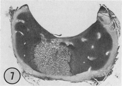
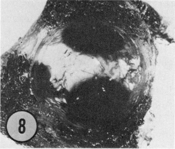
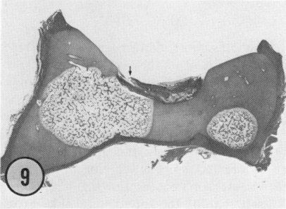
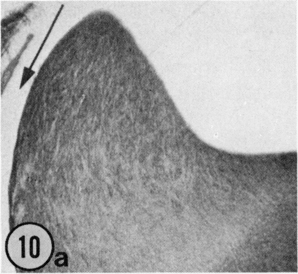
Seventy-four acetabula from a total of 140 normal human fetuses, obtained from abortions and deaths in the prenatal period, were used. The fetuses ranged from 9.1 to 40 cm in crown-rump length and are believed to be between 12 weeks and term. Acetabula were decalcified embedded in paraffin or celloidin, sectioned, and stained using conventional histologic techniques. Sections from the superior one-quarter of the acetabulum were examined for the initial appearance and later spread of osseous tissue. Throughout the fetal period bone was present only in the floor of the acetabulum and did not extend into the socket walls. Ossification was detected initially more posteriorly in the socket floor, and at all ages, ossification was more prominent on the ischial side of the socket. Despite the lack of osseous tissue a well-formed hyaline cartilage socket was present. The fetal labrum was composed of fibrous tissue with the density of fibers increasing with age. Typical-appearing chondrocytes were detected at only the inner articular margin of the labrum. Contributing from one-fifth to one-half of the socket depth, the labrum may play a greater role in containing the femoral head at birth than it does in the mature joint. In seven acetabula, from joints that were neither subluxated nor dislocated, an area of areolar tissue with capillaries was detected at the hyaline cartilage-labrum junction. Such defects may weaken the labrum and contribute to neonatal hip instability.
使用了从140例正常人类胎儿中获取的74个髋臼,这些胎儿是在产前因流产和死亡获得的。胎儿的顶臀长度在9.1至40厘米之间,据信处于12周龄至足月之间。髋臼经脱钙后包埋于石蜡或火棉胶中,切片,并采用传统组织学技术染色。检查髋臼上四分之一部分的切片,观察骨组织的初始出现情况及后续扩展情况。在整个胎儿期,骨仅存在于髋臼底部,未延伸至髋臼壁。骨化最初在髋臼底部更靠后的位置被检测到,并且在所有年龄段,髋臼坐骨侧的骨化更为明显。尽管缺乏骨组织,但仍存在一个发育良好的透明软骨髋臼。胎儿髋臼唇由纤维组织组成,纤维密度随年龄增加。仅在髋臼唇的内关节边缘检测到典型的软骨细胞。髋臼唇占髋臼深度的五分之一至二分之一,在出生时其在容纳股骨头方面可能比在成熟关节中发挥更大作用。在7个既未半脱位也未脱位的关节的髋臼中,在透明软骨 - 髋臼唇交界处检测到一个含有毛细血管的疏松组织区域。此类缺陷可能会削弱髋臼唇并导致新生儿髋关节不稳定。