• 文献检索
  • 文档翻译
  • 深度研究
  • 学术资讯
  • Suppr Zotero 插件Zotero 插件
  • 邀请有礼
  • 套餐&价格
  • 历史记录
应用&插件
Suppr Zotero 插件Zotero 插件浏览器插件Mac 客户端Windows 客户端微信小程序
定价
高级版会员购买积分包购买API积分包
服务
文献检索文档翻译深度研究API 文档MCP 服务
关于我们
关于 Suppr公司介绍联系我们用户协议隐私条款
关注我们

Suppr 超能文献

核心技术专利:CN118964589B侵权必究
粤ICP备2023148730 号-1Suppr @ 2026

文献检索

告别复杂PubMed语法,用中文像聊天一样搜索,搜遍4000万医学文献。AI智能推荐,让科研检索更轻松。

立即免费搜索

文件翻译

保留排版,准确专业,支持PDF/Word/PPT等文件格式,支持 12+语言互译。

免费翻译文档

深度研究

AI帮你快速写综述,25分钟生成高质量综述,智能提取关键信息,辅助科研写作。

立即免费体验

鹦鹉热衣原体在其发育周期中包膜的变化:互补复制品的冷冻断裂研究

Modifications of the envelope of Chlamydia psittaci during its developmental cycle: freeze-fracture study of complementary replicas.

作者信息

Louis C, Nicolas G, Eb F, Lefebvre J F, Orfila J

出版信息

J Bacteriol. 1980 Feb;141(2):868-75. doi: 10.1128/jb.141.2.868-875.1980.

DOI:10.1128/jb.141.2.868-875.1980
PMID:7364718
原文链接:https://pmc.ncbi.nlm.nih.gov/articles/PMC293698/
Abstract

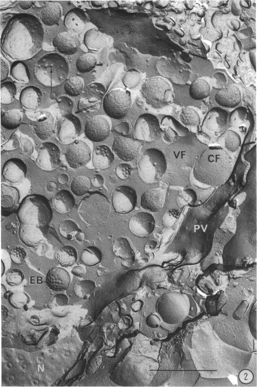
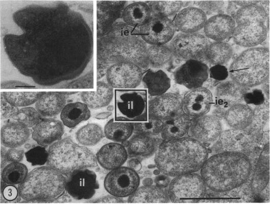
Examination of complementary replicas obtained by freeze-fracture of Chlamydia psittaci revealed, at the level of the plasma membrane, a progressive differentiation of "crate-like formations," which likely correspond to transmembranal pores. Recognition of "early" and "late" stages observed in the intermediate bodies permitted detailed study of the developmental cycle of this chlamydia.

摘要

对通过鹦鹉热衣原体冷冻断裂获得的互补复制品进行检查发现,在质膜水平上,“板条样结构”逐渐分化,这可能对应于跨膜孔。对中间体中观察到的“早期”和“晚期”阶段的识别使得能够对这种衣原体的发育周期进行详细研究。

https://cdn.ncbi.nlm.nih.gov/pmc/blobs/8737/293698/ef10c82899a6/jbacter00563-0443-a.jpg
https://cdn.ncbi.nlm.nih.gov/pmc/blobs/8737/293698/fc9c5c7671bf/jbacter00563-0439-a.jpg
https://cdn.ncbi.nlm.nih.gov/pmc/blobs/8737/293698/354844e5358f/jbacter00563-0440-a.jpg
https://cdn.ncbi.nlm.nih.gov/pmc/blobs/8737/293698/43edea02f67d/jbacter00563-0441-a.jpg
https://cdn.ncbi.nlm.nih.gov/pmc/blobs/8737/293698/6e2765d33b6e/jbacter00563-0442-a.jpg
https://cdn.ncbi.nlm.nih.gov/pmc/blobs/8737/293698/ef10c82899a6/jbacter00563-0443-a.jpg

相似文献

1
Modifications of the envelope of Chlamydia psittaci during its developmental cycle: freeze-fracture study of complementary replicas.鹦鹉热衣原体在其发育周期中包膜的变化:互补复制品的冷冻断裂研究
J Bacteriol. 1980 Feb;141(2):868-75. doi: 10.1128/jb.141.2.868-875.1980.
2
Surface projections of Chlamydia psittaci elementary bodies as revealed by freeze-deep-etching.鹦鹉热衣原体原体的表面投影通过冷冻深度蚀刻显示。
J Bacteriol. 1982 Aug;151(2):1040-2. doi: 10.1128/jb.151.2.1040-1042.1982.
3
[The replication cycle of Chlamydia trachomatis and Chlamydia psittaci: ultrastructural analysis].[沙眼衣原体和鹦鹉热衣原体的复制周期:超微结构分析]
Boll Ist Sieroter Milan. 1979 Nov 30;58(5):395-405.
4
Electron microscopic observations of surface projections on Chlamydia psittaci reticulate bodies.鹦鹉热衣原体网状体表面突起的电子显微镜观察
J Bacteriol. 1982 Apr;150(1):358-64. doi: 10.1128/jb.150.1.358-364.1982.
5
Arrays of hemispheric surface projections on Chlamydia psittaci and Chlamydia trachomatis observed by scanning electron microscopy.通过扫描电子显微镜观察到的鹦鹉热衣原体和沙眼衣原体上的半球形表面突起阵列。
J Bacteriol. 1979 Apr;138(1):241-4. doi: 10.1128/jb.138.1.241-244.1979.
6
Chlamydia psittaci: inclusion vacuole ultrastructure.
Can J Microbiol. 1980 Mar;26(3):396-401. doi: 10.1139/m80-065.
7
Observations of the surface projections of infectious small cell of Chlamydia psittaci in thin sections.鹦鹉热衣原体感染性小细胞在超薄切片中的表面投影观察。
J Electron Microsc (Tokyo). 1976;25(3):169-70.
8
Synthesis of disulfide-bonded outer membrane proteins during the developmental cycle of Chlamydia psittaci and Chlamydia trachomatis.鹦鹉热衣原体和沙眼衣原体发育周期中双硫键结合外膜蛋白的合成
J Bacteriol. 1986 Feb;165(2):379-85. doi: 10.1128/jb.165.2.379-385.1986.
9
The effect of purification on the ultrastructure and infectivity of egg-attenuated Chlamydia psittaci (6BC).纯化对减毒鸡衣原体(6BC)卵内超微结构和感染性的影响。
Can J Microbiol. 1975 Oct;21(10):1448-63. doi: 10.1139/m75-216.
10
Surface projections and internal structure of Chlamydia psittaci.鹦鹉热衣原体的体表投影及内部结构
J Bacteriol. 1978 Mar;133(3):1514-6. doi: 10.1128/jb.133.3.1514-1516.1978.

引用本文的文献

1
Pathogen-host reorganization during Chlamydia invasion revealed by cryo-electron tomography.冷冻电子断层扫描揭示衣原体入侵过程中的病原体-宿主重组
Cell Microbiol. 2014 Oct;16(10):1457-72. doi: 10.1111/cmi.12310. Epub 2014 Jun 3.
2
Significance of host cell kinesin in the development of Chlamydia psittaci.宿主细胞驱动蛋白在鹦鹉热衣原体发育中的意义。
Infect Immun. 1999 Oct;67(10):5441-6. doi: 10.1128/IAI.67.10.5441-5446.1999.
3
Surface projections of a chlamydia-like parasite of midge larvae.蠓幼虫类衣原体寄生虫的体表投影

本文引用的文献

1
Electron Microscopic Observations on the Fine Structure of Cell Walls of Chlamydia psittaci.鹦鹉热衣原体细胞壁精细结构的电子显微镜观察
J Bacteriol. 1970 Dec;104(3):1332-7. doi: 10.1128/jb.104.3.1332-1337.1970.
2
Structure of purified cell walls of dense forms of meningopneumonitis organisms.脑膜肺炎病菌致密形态的纯化细胞壁结构
J Bacteriol. 1966 Jan;91(1):409-13. doi: 10.1128/jb.91.1.409-413.1966.
3
[Study with electron microscopy of the development and morphology of Chlamydia psittaci in mouse macrophages].
J Bacteriol. 1980 Dec;144(3):1174-5. doi: 10.1128/jb.144.3.1174-1175.1980.
4
Identification of a major envelope protein in Chlamydia spp.衣原体属中一种主要包膜蛋白的鉴定
J Bacteriol. 1981 Apr;146(1):426-9. doi: 10.1128/jb.146.1.426-429.1981.
5
Surface projections of Chlamydia psittaci elementary bodies as revealed by freeze-deep-etching.鹦鹉热衣原体原体的表面投影通过冷冻深度蚀刻显示。
J Bacteriol. 1982 Aug;151(2):1040-2. doi: 10.1128/jb.151.2.1040-1042.1982.
6
Structural and polypeptide differences between envelopes of infective and reproductive life cycle forms of Chlamydia spp.衣原体属感染性和繁殖性生命周期形式的包膜之间的结构和多肽差异
J Bacteriol. 1984 Jan;157(1):13-20. doi: 10.1128/jb.157.1.13-20.1984.
7
Isolation and electron microscopic observations of intracytoplasmic inclusions containing Chlamydia psittaci.鹦鹉热衣原体胞质内包涵体的分离及电镜观察
J Bacteriol. 1981 Jan;145(1):605-12. doi: 10.1128/jb.145.1.605-612.1981.
8
Recent advances in Chlamydia trachomatis.沙眼衣原体的最新进展
Eur J Epidemiol. 1985 Dec;1(4):235-56. doi: 10.1007/BF00237099.
9
New view of the surface projections of Chlamydia trachomatis.沙眼衣原体表面投影的新观点。
J Bacteriol. 1985 Oct;164(1):344-9. doi: 10.1128/jb.164.1.344-349.1985.
10
Disulfide-mediated interactions of the chlamydial major outer membrane protein: role in the differentiation of chlamydiae?衣原体主要外膜蛋白的二硫键介导的相互作用:在衣原体分化中的作用?
J Bacteriol. 1985 Jan;161(1):25-31. doi: 10.1128/jb.161.1.25-31.1985.
Ann Inst Pasteur (Paris). 1970 Aug;119(2):222-31.
4
Fracture faces of frozen membranes.冷冻膜的断裂面
Proc Natl Acad Sci U S A. 1966 May;55(5):1048-56. doi: 10.1073/pnas.55.5.1048.
5
[Comparative ultrastructure of Ricketsiella melolonthae (Rickettsiales Wolbachiae) and of the agent of venereal lymphogranulomatosis (Rickettsiales Clamydiaceae)].
C R Acad Hebd Seances Acad Sci D. 1971 Jun 7;272(23):2972-4.
6
Fracture faces in the cell envelope of Escherichia coli.大肠杆菌细胞膜中的断裂面。
J Bacteriol. 1971 Oct;108(1):474-81. doi: 10.1128/jb.108.1.474-481.1971.
7
[Ultrastructure and cytochemistry of Chlamydia Psittaci].[鹦鹉热衣原体的超微结构与细胞化学]
Arch Gesamte Virusforsch. 1971;33(3):271-80.
8
[Fine structure of trachoma agent as observed by freeze-etching electron microscopy (author's transl)].[通过冷冻蚀刻电子显微镜观察到的沙眼病原体的精细结构(作者译)]
Nippon Ganka Gakkai Zasshi. 1973 Sep;77(9):1184-93.
9
Structure and function of the cell envelope of gram-negative bacteria.革兰氏阴性菌细胞包膜的结构与功能
Bacteriol Rev. 1974 Mar;38(1):87-110. doi: 10.1128/br.38.1.87-110.1974.
10
The fluid mosaic model of the structure of cell membranes.细胞膜结构的流动镶嵌模型。
Science. 1972 Feb 18;175(4023):720-31. doi: 10.1126/science.175.4023.720.