• 文献检索
  • 文档翻译
  • 深度研究
  • 学术资讯
  • Suppr Zotero 插件Zotero 插件
  • 邀请有礼
  • 套餐&价格
  • 历史记录
应用&插件
Suppr Zotero 插件Zotero 插件浏览器插件Mac 客户端Windows 客户端微信小程序
定价
高级版会员购买积分包购买API积分包
服务
文献检索文档翻译深度研究API 文档MCP 服务
关于我们
关于 Suppr公司介绍联系我们用户协议隐私条款
关注我们

Suppr 超能文献

核心技术专利:CN118964589B侵权必究
粤ICP备2023148730 号-1Suppr @ 2026

文献检索

告别复杂PubMed语法,用中文像聊天一样搜索,搜遍4000万医学文献。AI智能推荐,让科研检索更轻松。

立即免费搜索

文件翻译

保留排版,准确专业,支持PDF/Word/PPT等文件格式,支持 12+语言互译。

免费翻译文档

深度研究

AI帮你快速写综述,25分钟生成高质量综述,智能提取关键信息,辅助科研写作。

立即免费体验

兔唇黏膜上皮中默克尔细胞的分化

Merkel cell differentiation in the labial mucous epithelium of the rabbit.

作者信息

Tachibana T, Nawa T

出版信息

J Anat. 1980 Aug;131(Pt 1):145-55.

PMID:7440398
原文链接:https://pmc.ncbi.nlm.nih.gov/articles/PMC1233293/
Abstract

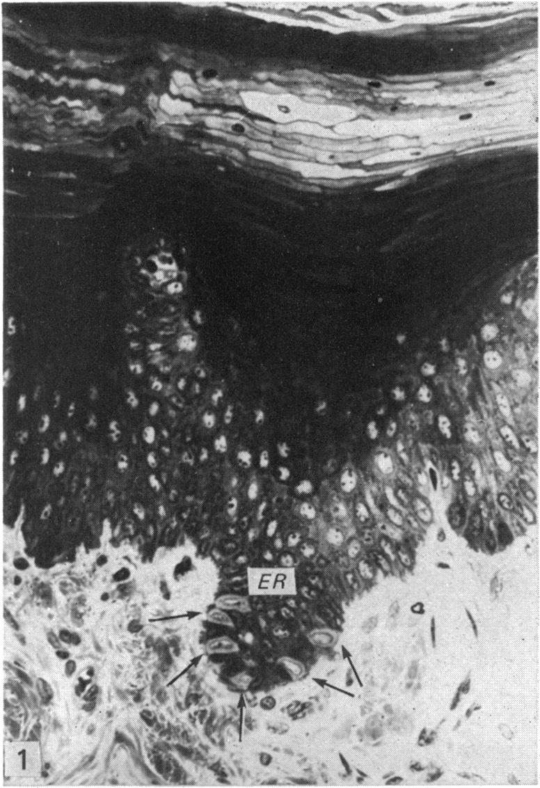
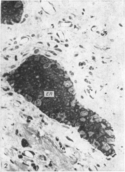
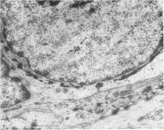
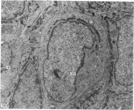
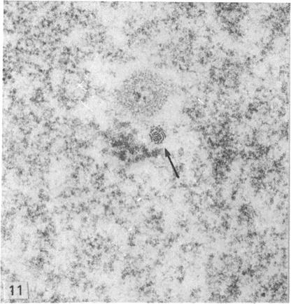
To clarify the relation between the so-called 'transitional' cell and the Merkel cell, the frequency of these cells in the labial mucosal epithelium was compared between infant, juvenile and adult rabbits. The transitional cell was observed in serial ultrathin sections. Transitional cells were most numerous in the infant and juvenile, while the definitive Merkel cell showed a reciprocal increase in number. In the infant and juvenile, the transitional cells differed slightly from each other in their ultrastructure: some resembled immature keratinocytes, others resembled definitive Merkel cells. All transitional cells contained the Merkel cell granules and the intranuclear rodlet. It was interpreted that the Merkel cell in the labial mucous epithelium develops from the transitional cell. Neither definitive Merkel cells not transitional cells were identified in the lamina propria. The present circumstantial evidence favours the hypothesis that Merkel cells differentiate from precursors in the basal layer of the epithelium.

摘要

为阐明所谓的“过渡性”细胞与默克尔细胞之间的关系,比较了婴儿、幼年和成年兔唇黏膜上皮中这些细胞的出现频率。在连续超薄切片中观察到了过渡性细胞。过渡性细胞在婴儿和幼年兔中数量最多,而成熟的默克尔细胞数量则呈相反增加。在婴儿和幼年兔中,过渡性细胞的超微结构彼此略有不同:一些类似于未成熟的角质形成细胞,另一些类似于成熟的默克尔细胞。所有过渡性细胞均含有默克尔细胞颗粒和核内小杆。据推测,唇黏膜上皮中的默克尔细胞由过渡性细胞发育而来。在固有层中未发现成熟的默克尔细胞和过渡性细胞。目前的间接证据支持默克尔细胞由上皮基底层的前体细胞分化而来这一假说。

https://cdn.ncbi.nlm.nih.gov/pmc/blobs/89c1/1233293/55c318596f6f/janat00229-0155-c.jpg
https://cdn.ncbi.nlm.nih.gov/pmc/blobs/89c1/1233293/9c3ea1044fe4/janat00229-0149-a.jpg
https://cdn.ncbi.nlm.nih.gov/pmc/blobs/89c1/1233293/b84fc7200cfe/janat00229-0149-b.jpg
https://cdn.ncbi.nlm.nih.gov/pmc/blobs/89c1/1233293/ea86d53702fc/janat00229-0149-c.jpg
https://cdn.ncbi.nlm.nih.gov/pmc/blobs/89c1/1233293/30edbb57a722/janat00229-0151-a.jpg
https://cdn.ncbi.nlm.nih.gov/pmc/blobs/89c1/1233293/def2d6c975a9/janat00229-0151-b.jpg
https://cdn.ncbi.nlm.nih.gov/pmc/blobs/89c1/1233293/f97c3bd68db2/janat00229-0152-a.jpg
https://cdn.ncbi.nlm.nih.gov/pmc/blobs/89c1/1233293/f1787c47fa83/janat00229-0152-b.jpg
https://cdn.ncbi.nlm.nih.gov/pmc/blobs/89c1/1233293/d0cc6429d277/janat00229-0154-a.jpg
https://cdn.ncbi.nlm.nih.gov/pmc/blobs/89c1/1233293/288332c47983/janat00229-0154-b.jpg
https://cdn.ncbi.nlm.nih.gov/pmc/blobs/89c1/1233293/a773ee3299b9/janat00229-0155-a.jpg
https://cdn.ncbi.nlm.nih.gov/pmc/blobs/89c1/1233293/69065ca3ef3c/janat00229-0155-b.jpg
https://cdn.ncbi.nlm.nih.gov/pmc/blobs/89c1/1233293/55c318596f6f/janat00229-0155-c.jpg

相似文献

1
Merkel cell differentiation in the labial mucous epithelium of the rabbit.兔唇黏膜上皮中默克尔细胞的分化
J Anat. 1980 Aug;131(Pt 1):145-55.
2
[Melanocytes, Langerhans and Merkel cells in oral epithelium].[口腔上皮中的黑素细胞、朗格汉斯细胞和默克尔细胞]
Acta Otolaryngol. 1975 Sep-Oct;80(3-4):301-11.
3
Ultrastructure of Merkel cells in the hard palate of the squirrel monkey (Saimiri sciureus).松鼠猴(Saimiri sciureus)硬腭中默克尔细胞的超微结构。
Am J Anat. 1980 Feb;157(2):155-67. doi: 10.1002/aja.1001570204.
4
Migration of Merkel cells in the labial mucous epithelium of adult rabbits following mental nerve resection.颏神经切除术后成年兔唇黏膜上皮中默克尔细胞的迁移
Cell Tissue Res. 1982;223(3):659-64. doi: 10.1007/BF00218484.
5
An experimental study of the influence of sensory nerve fibers on Merkel cell differentiation in the labial mucosa of the rabbits.感觉神经纤维对兔唇黏膜默克尔细胞分化影响的实验研究
Arch Histol Jpn. 1983 Sep;46(4):469-77. doi: 10.1679/aohc.46.469.
6
Ultrastructure of Merkel corpuscles and so-called "transitional" cells in the white Leghorn chicken.白来航鸡中默克尔小体及所谓“过渡性”细胞的超微结构
Am J Anat. 1978 Apr;151(4):453-73. doi: 10.1002/aja.1001510402.
7
Ultrastructural study of rabbit buccal epithelial cells and intercellular junction by scanning and transmission electron microscopy.
J Electron Microsc (Tokyo). 1993 Dec;42(6):389-93.
8
Merkel cell development in the wound healing in the labial mucosa of adult rabbits.成年兔唇黏膜伤口愈合过程中的默克尔细胞发育
Arch Histol Jpn. 1981 May;44(2):151-65. doi: 10.1679/aohc1950.44.151.
9
The Merkel cell in oral human mucosa.口腔人类黏膜中的默克尔细胞。
J Dent Res. 1977 Nov;56(11):1303-12. doi: 10.1177/00220345770560110201.
10
Effects of collagen concentration on epithelial differentiation: in vitro analysis with a mucosal tissue model.胶原蛋白浓度对上皮分化的影响:使用黏膜组织模型进行的体外分析
J Craniofac Genet Dev Biol. 1993 Apr-Jun;13(2):127-33.

引用本文的文献

1
Dendritic atoh1a+ cells serve as Merkel cell precursors during skin development and regeneration.在皮肤发育和再生过程中,树突状的atoh1a+细胞充当默克尔细胞前体。
Development. 2025 Oct 15;152(20). doi: 10.1242/dev.204810. Epub 2025 Jun 20.
2
Immunohistochemical analyses point to epidermal origin of human Merkel cells.免疫组织化学分析表明人类默克尔细胞起源于表皮。
Histochem Cell Biol. 2014 Apr;141(4):407-21. doi: 10.1007/s00418-013-1168-8. Epub 2013 Nov 30.
3
Epidermal progenitors give rise to Merkel cells during embryonic development and adult homeostasis.

本文引用的文献

1
The intraepidermal innervation of the snout skin of the opossum. A light and electron microscope study, with observations on the nature of Merkel's Tastzellen.负鼠口鼻部皮肤的表皮内神经支配。一项光镜和电镜研究,兼对默克尔味觉细胞性质的观察。
J Cell Biol. 1965 Jul;26(1):79-97. doi: 10.1083/jcb.26.1.79.
2
Fine structures of Merkel cells and associated nerve fibers in the epidermis of certain mammalian species.某些哺乳动物物种表皮中默克尔细胞及相关神经纤维的精细结构。
Arch Histol Jpn. 1969 Apr;30(3):295-313. doi: 10.1679/aohc1950.30.295.
3
The structure and function of the Haarscheibe.
在胚胎发育和成年期稳态过程中,表皮祖细胞产生默克尔细胞。
J Cell Biol. 2009 Oct 5;187(1):91-100. doi: 10.1083/jcb.200907080. Epub 2009 Sep 28.
4
Proliferative Merkel cells were not detected in human skin.在人类皮肤中未检测到增殖性默克尔细胞。
Arch Dermatol Res. 1996 Apr;288(4):184-7. doi: 10.1007/BF02505222.
5
Migration of Merkel cells in the labial mucous epithelium of adult rabbits following mental nerve resection.颏神经切除术后成年兔唇黏膜上皮中默克尔细胞的迁移
Cell Tissue Res. 1982;223(3):659-64. doi: 10.1007/BF00218484.
6
The development of the Merkel cells in the tentacles of Xenopus laevis larvae.非洲爪蟾幼虫触须中默克尔细胞的发育。
Anat Embryol (Berl). 1987;176(4):493-500. doi: 10.1007/BF00310089.
7
Merkel cell mitoses in vibrissae: an ultrastructural study.触须中默克尔细胞的有丝分裂:一项超微结构研究。
J Anat. 1987 Aug;153:241-4.
8
Morphology and immunocytochemistry of two endocrine cell types in the guinea-pig esophageal epithelium.
Cell Tissue Res. 1989 Jun;256(3):635-43. doi: 10.1007/BF00225614.
环层小体的结构与功能。
J Comp Neurol. 1967 Dec;131(4):459-74. doi: 10.1002/cne.901310406.
4
Ultrastructural observations on Merkel cells in human foetal skin.人胎儿皮肤中默克尔细胞的超微结构观察
J Anat. 1970 Mar;106(Pt 2):411.
5
The Herman Beerman lecture: embryology of human skin, a review of ultrastructural studies.
J Invest Dermatol. 1971 Sep;57(3):133-43. doi: 10.1111/1523-1747.ep12261482.
6
Fine structure of Merkel cell in human oral mucosa.人口腔黏膜中默克尔细胞的精细结构。
J Invest Dermatol. 1972 Jun;58(6):381-7. doi: 10.1111/1523-1747.ep12540607.
7
The ultrastructure of the skin of human embryos. X. Merkel tactile cells in the finger and nail.人类胚胎皮肤的超微结构。X. 手指和指甲中的默克尔触觉细胞。
J Anat. 1972 Jan;111(Pt 1):99-120.
8
The structure and function of a slowly adapting touch corpuscle in hairy skin.有毛皮肤中慢适应性触觉小体的结构与功能。
J Physiol. 1969 Feb;200(3):763-96. doi: 10.1113/jphysiol.1969.sp008721.
9
Cell types in cutaneous type I mechanoreceptors (Haarscheiben) and their alterations with injury.皮肤I型机械感受器(Haarscheiben)中的细胞类型及其损伤后的改变。
Am J Anat. 1974 Sep;141(1):105-26. doi: 10.1002/aja.1001410107.
10
A Merkel cell nuclear inclusion.一个默克尔细胞核内包涵体。
J Invest Dermatol. 1973 Dec;61(6):334-8. doi: 10.1111/1523-1747.ep12676616.