Faussone Pellegrini M S, Manneschi L I, Manneschi L
Department of Human Anatomy and Histology, University of Florence, Italy.
Gut. 1995 Oct;37(4):493-8. doi: 10.1136/gut.37.4.493.
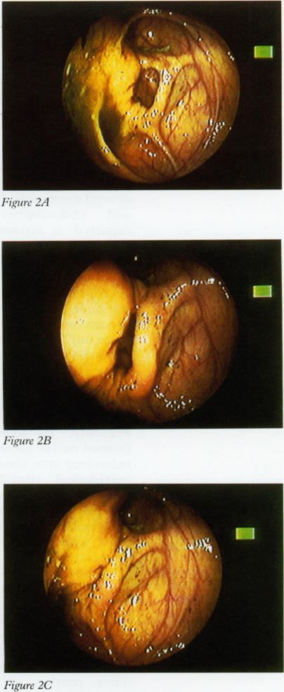
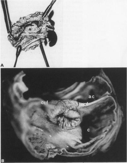
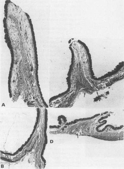
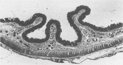
Sphincteric anatomy and function are present at the caecocolonic junction in several mammals. In humans, radiologists and endoscopists have respectively reported a circumferential contraction and a prominent ileocaecal fold at the border area between the caecum and the ascending colon. Anatomical findings on necropsy material failed to confirm its presence. Microscopic studies on surgical specimens showed the existence of muscular and innervational patterns different from those of adjacent areas. The aim of this work was to confirm the existence of a specialised fold at the caecocolonic junction in humans and to ascertain its role by carrying out a study of functional anatomy. Pancolonoscopies were performed on 100 patients and ileocaecal fold behaviour was observed before and after mechanical stimulation. Isolated ileocaecocolonic regions, surgically obtained, were filled with a fixative solution to study their macro and microscopic morphology after stimulation. Endoscopically, the ileocaecal fold was semilunar or circular in shape and spontaneous or evoked spasms occurred in 52 patients. A prominent circular fold could be seen in surgical specimens after stimulation. The entire muscle coat deeply penetrated this fold, showing the features characteristic of the ileocaecal junction. In particular, the inner portion of the circular muscle showed a peculiar arrangement and was thicker than elsewhere. These results show that in humans the caecocolonic junction is provided with a sphincter morphology and function. Little is known about its physiological relevance in ileal flow accommodation and caecal filling and emptying but it should not be underestimated with regard to some colonic motility disorders.
在几种哺乳动物的盲结肠交界处存在括约肌的解剖结构和功能。在人类中,放射科医生和内镜医生分别报告了在盲肠和升结肠之间的边界区域存在环形收缩和明显的回盲皱襞。尸检材料的解剖学发现未能证实其存在。对手术标本的显微镜研究显示,其肌肉和神经支配模式与相邻区域不同。这项工作的目的是通过进行功能解剖学研究来证实人类盲结肠交界处存在特殊皱襞,并确定其作用。对100名患者进行了全结肠镜检查,并观察了机械刺激前后回盲皱襞的行为。手术获取的孤立回盲结肠区域用固定液填充,以研究刺激后的大体和微观形态。在内镜下,回盲皱襞呈半月形或圆形,52例患者出现自发或诱发的痉挛。刺激后在手术标本中可见明显的环形皱襞。整个肌层深深穿透该皱襞,呈现出回盲交界处的特征。特别是,环形肌的内部呈现出特殊的排列,且比其他部位更厚。这些结果表明,在人类中,盲结肠交界处具有括约肌的形态和功能。关于其在回肠液流调节以及盲肠充盈和排空方面的生理相关性知之甚少,但在某些结肠动力障碍方面不应被低估。