Takemura R, Okabe S, Umeyama T, Hirokawa N
Department of Anatomy and Cell Biology, School of Medicine, University of Tokyo, Japan.
Mol Biol Cell. 1995 Aug;6(8):981-96. doi: 10.1091/mbc.6.8.981.
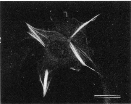
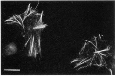
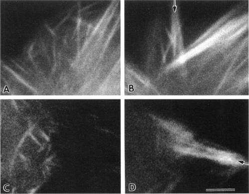
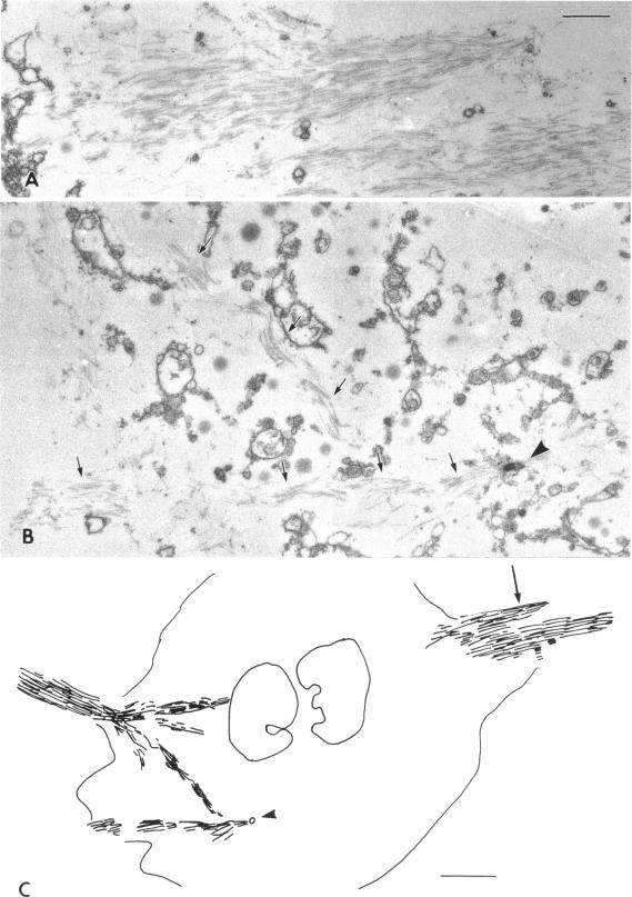
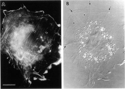
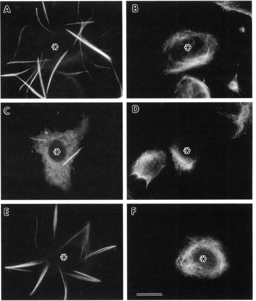
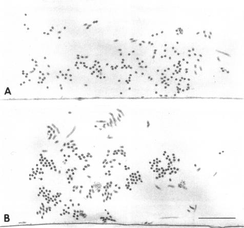
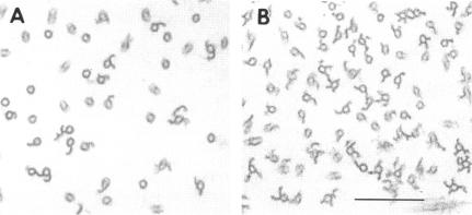
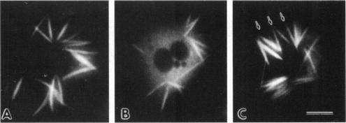
Microtubule bundles reminiscent of those found in neuronal processes are formed in fibroblasts and Sf9 cells that are transfected with the microtubule-associated proteins tau, MAP2, or MAP2c. To analyze the assembly process of these bundles and its relation to the microtubule polarity, we depolymerized the bundles formed in MAP2c-transfected COS cells using nocodazole, and observed the process of assembly of microtubule bundles after removal of the drug in cells microinjected with rhodamine-labeled tubulin. Within minutes of its removal, numerous short microtubule fragments were observed throughout the cytoplasm. These short fragments were randomly oriented and were already bundled. Somewhat longer, but still short bundles, were then found in the peripheral cytoplasm. These bundles became the primordium of the larger bundles, and gradually grew in length and width. The polarity orientation of microtubules in the reformed bundle as determined by "hook" procedure using electron microscope was uniform with the plus end distal to the cell nucleus. The results suggest that some mechanism(s) exists to orient the polarity of microtubules, which are not in direct continuity with the centrosome, during the formation of large bundles. The observed process presents a useful model system for studying the organization of microtubules that are not directly associated with the centrosomes, such as those observed in axons.
在转染了微管相关蛋白tau、MAP2或MAP2c的成纤维细胞和Sf9细胞中,形成了类似于在神经元突起中发现的微管束。为了分析这些微管束的组装过程及其与微管极性的关系,我们用诺考达唑使在转染了MAP2c的COS细胞中形成的微管束解聚,并在显微注射了罗丹明标记微管蛋白的细胞中去除药物后观察微管束的组装过程。在去除药物后的几分钟内,在整个细胞质中观察到大量短的微管片段。这些短片段随机取向且已经成束。然后在外周细胞质中发现了稍长但仍然较短的微管束。这些微管束成为较大微管束的原基,并逐渐在长度和宽度上生长。使用电子显微镜通过“钩”程序确定的重新形成的微管束中微管的极性取向是一致的,正端远离细胞核。结果表明,在大微管束形成过程中,存在某种机制来定向与中心体没有直接连续性的微管的极性。观察到的过程为研究与中心体没有直接关联的微管的组织提供了一个有用的模型系统,例如在轴突中观察到的微管。