Sim L J, Selley D E, Childers S R
Department of Physiology and Pharmacology, Bowman Gray School of Medicine, Wake Forest University, Winston-Salem, NC 27157, USA.
Proc Natl Acad Sci U S A. 1995 Aug 1;92(16):7242-6. doi: 10.1073/pnas.92.16.7242.
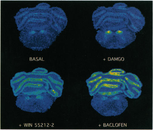
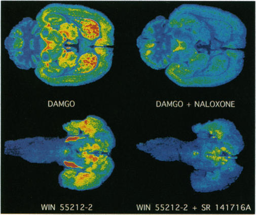
Agonists stimulate guanylyl 5'-[gamma-[35S]thio]-triphosphate (GTP[gamma-35S]) binding to receptor-coupled guanine nucleotide binding protein (G proteins) in cell membranes as revealed in the presence of excess GDP. We now report that this reaction can be used to neuroanatomically localize receptor-activated G proteins in brain sections by in vitro autoradiography of GTP[gamma-35S] binding. Using the mu opioid-selective peptide [D-Ala2,N-MePhe4,Gly5-ol]enkephalin (DAMGO) as an agonist in rat brain sections and isolated thalamic membranes, agonist stimulation of GTP[gamma-35S] binding required the presence of excess GDP (1-2 mM GDP in sections vs. 10-30 microM GDP in membranes) to decrease basal G-protein activity and reveal agonist-stimulated GTP[gamma-35S] binding. Similar concentrations of DAMGO were required to stimulate GTP[gamma-35S] binding in sections and membranes. To demonstrate the general applicability of the technique, agonist-stimulated GTP[gamma-35S] binding in tissue sections was assessed with agonists for the mu opioid (DAMGO), cannabinoid (WIN 55212-2), and gamma-aminobutyric acid type B (baclofen) receptors. For opioid and cannabinoid receptors, agonist stimulation of GTP[gamma-35S] binding was blocked by incubation with agonists in the presence of the appropriate antagonists (naloxone for mu opioid and SR-141716A for cannabinoid), thus demonstrating that the effect was specifically receptor mediated. The anatomical distribution of agonist-stimulated GTP[gamma-35S] binding qualitatively paralleled receptor distribution as determined by receptor binding autoradiography. However, quantitative differences suggest that variations in coupling efficiency may exist between different receptors in various brain regions. This technique provides a method of functional neuroanatomy that identifies changes in the activation of G proteins by specific receptors.
在过量GDP存在的情况下,激动剂可刺激鸟苷酰5'-[γ-[35S]硫代] -三磷酸(GTP[γ-35S])与细胞膜中受体偶联的鸟嘌呤核苷酸结合蛋白(G蛋白)结合。我们现在报告,该反应可通过GTP[γ-35S]结合的体外放射自显影,在脑切片中对受体激活的G蛋白进行神经解剖定位。在大鼠脑切片和分离的丘脑膜中,使用μ阿片受体选择性肽[D-Ala2,N-MePhe4,Gly5-ol]脑啡肽(DAMGO)作为激动剂,激动剂刺激GTP[γ-35S]结合需要存在过量的GDP(切片中为1-2 mM GDP,而膜中为10-30 μM GDP)以降低基础G蛋白活性并揭示激动剂刺激的GTP[γ-35S]结合。在切片和膜中刺激GTP[γ-35S]结合需要相似浓度的DAMGO。为了证明该技术的普遍适用性,用μ阿片受体(DAMGO)、大麻素受体(WIN 55212-2)和γ-氨基丁酸B型受体(巴氯芬)的激动剂评估组织切片中激动剂刺激的GTP[γ-35S]结合。对于阿片受体和大麻素受体,在适当拮抗剂(μ阿片受体用纳洛酮,大麻素受体用SR-141716A)存在下与激动剂一起孵育可阻断激动剂对GTP[γ-35S]结合的刺激,从而证明该效应是由受体特异性介导的。激动剂刺激的GTP[γ-35S]结合的解剖分布在定性上与通过受体结合放射自显影确定的受体分布平行。然而,定量差异表明不同脑区的不同受体之间可能存在偶联效率的差异。该技术提供了一种功能性神经解剖学方法,可识别特定受体对G蛋白激活的变化。