Brown W C, Zhao S, Woods V M, Tripp C A, Tetzlaff C L, Heussler V T, Dobbelaere D A, Rice-Ficht A C
Department of Veterinary Pathobiology, Texas A&M University, College Station 77843.
Infect Immun. 1993 Jan;61(1):236-44. doi: 10.1128/iai.61.1.236-244.1993.
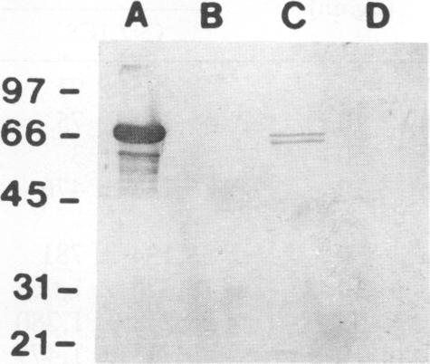
Previous studies have demonstrated the serologic and T-cell immunogenicity for cattle of a recombinant form of the apical complex-associated 77-kDa merozite protein of Babesia bovis, designated Bb-1. The present study characterizes the immunogenic epitopes of the Bb-1 protein. A series of recombinant truncated fusion proteins spanning the majority of the Bb-1 protein were expressed in Escherichia coli, and their reactivities with bovine peripheral blood mononuclear cells and T-cell clones derived from B. bovis-immune cattle and with rabbit antibodies were determined. Lymphocytes from two immune cattle were preferentially stimulated by the N-terminal half of the Bb-1 protein (amino acids 23 to 266, termed Bb-1A), localizing the T-cell epitopes to the Bb-1A portion of the molecule. CD4+ T-cell clones derived by stimulation with the intact Bb-1 fusion protein were used to identify two T-cell epitopes in the Bb-1A protein, consisting of amino acids SVVLLSAFSGN VWANEAEVSQVVK and FSDVDKTKSTEKT (residues 23 to 46 and 82 to 94). In contrast, rabbit antiserum raised against the intact fusion protein reacted only with the C-terminal half of the protein (amino acids 267 to 499, termed Bb-1B), which contained 28 tandem repeats of the tetrapeptide PAEK or PAET. Biological assays and Northern (RNA) blot analyses for cytokines revealed that following activation with concanavalin A, T-cell clones reactive against the two Bb-1A epitopes produced interleukin-2, gamma interferon, and tumor necrosis factors beta and alpha, but not interleukin-4, suggesting that the Bb-1 antigen preferentially stimulates the Th1 subset of CD4+ T cells in cattle. The studies described here report for the first time the characterization, by cytokine production, of the Th1 subset of bovine T cells and show that, as in mice, protozoal antigens can induce Th1 cells in ruminants. This first demonstration of B. bovis-encoded Th1 cell epitopes provides a rationale for incorporation of all or part of the Bb-1 protein into a recombinant vaccine.
先前的研究已证明,一种重组形式的牛巴贝斯虫顶端复合体相关的77 kDa裂殖子蛋白(命名为Bb-1)对牛具有血清学和T细胞免疫原性。本研究对Bb-1蛋白的免疫原性表位进行了表征。一系列跨越Bb-1蛋白大部分区域的重组截短融合蛋白在大肠杆菌中表达,并测定了它们与牛外周血单个核细胞、源自牛巴贝斯虫免疫牛的T细胞克隆以及兔抗体的反应性。来自两头免疫牛的淋巴细胞优先受到Bb-1蛋白N端一半(氨基酸23至266,称为Bb-1A)的刺激,从而将T细胞表位定位到该分子的Bb-1A部分。用完整的Bb-1融合蛋白刺激产生的CD4+ T细胞克隆用于鉴定Bb-1A蛋白中的两个T细胞表位,分别由氨基酸SVVLLSAFSGN VWANEAEVSQVVK和FSDVDKTKSTEKT(第23至46位和第82至94位残基)组成。相比之下,针对完整融合蛋白产生的兔抗血清仅与该蛋白的C端一半(氨基酸267至499,称为Bb-1B)发生反应,该部分含有28个四肽PAEK或PAET的串联重复序列。细胞因子的生物学检测和Northern(RNA)印迹分析显示,在用伴刀豆球蛋白A激活后,对两个Bb-1A表位有反应的T细胞克隆产生白细胞介素-2、γ干扰素以及肿瘤坏死因子β和α,但不产生白细胞介素-4,这表明Bb-1抗原优先刺激牛体内CD4+ T细胞的Th1亚群。此处描述的研究首次通过细胞因子产生对牛T细胞的Th1亚群进行了表征,并表明,与小鼠一样,原生动物抗原可在反刍动物中诱导Th1细胞。首次证明牛巴贝斯虫编码的Th1细胞表位为将全部或部分Bb-1蛋白纳入重组疫苗提供了理论依据。