Thomas G J, Benevides J M, Overman S A, Ueda T, Ushizawa K, Saitoh M, Tsuboi M
Division of Cell Biology and Biophysics, School of Biological Sciences, University of Missouri-Kansas City 64110, USA.
Biophys J. 1995 Mar;68(3):1073-88. doi: 10.1016/S0006-3495(95)80282-1.
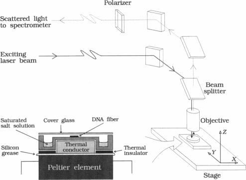
Polarized Raman spectra of oriented fibers of calf thymus DNA in the A and B conformations have been obtained by use of a Raman microscope operating in the 180 degrees back-scattering geometry. The following polarized Raman intensities in the spectral interval 200-1800 cm-1 were measured with both 514.5 and 488.0 nm laser excitations: (1) Icc, in which the incident and scattered light are polarized parallel to the DNA helical axis (c axis); (2) Ibb, in which the incident and scattered light are polarized perpendicular to c; and (3) Ibc and Icb, in which the incident and scattered light are polarized in mutually perpendicular directions. High degrees of structural homogeneity and unidirectional orientation were confirmed for both the A and B form fibers, as judged by comparison of the observed Raman markers and intensity anisotropies with measurements reported previously for oligonucleotide single crystals of known three-dimensional structures. The fiber Raman anisotropies have been combined with solution Raman depolarization ratios to evaluate the local tensors corresponding to key conformation-sensitive Raman bands of the DNA bases and sugar-phosphate backbone. The present study yields novel vibrational assignments for both A DNA and BDNA conformers and also confirms many previously proposed Raman vibrational assignments. Among the significant new findings are the demonstration of complex patterns of A form and B form indicator bands in the spectral intervals 750-900 and 1050-1100 cm-1, the identification of highly anisotropic tensors corresponding to vibrations of base, deoxyribose, and phosphate moieties, and the determination of relatively isotropic Raman tensors for the symmetrical stretching mode of phosphodioxy groups in A and B DNA. The present fiber results provide a basis for exploitation of polarized Raman spectroscopy to determine DNA helix orientation as well as to probe specific nucleotide residue orientations in nucleoproteins, viruses, and other complex biological assemblies.
利用在180度背散射几何构型下工作的拉曼显微镜,获得了处于A和B构象的小牛胸腺DNA取向纤维的偏振拉曼光谱。在200 - 1800 cm-1光谱区间内,使用514.5和488.0 nm激光激发测量了以下偏振拉曼强度:(1)Icc,其中入射光和散射光平行于DNA螺旋轴(c轴)偏振;(2)Ibb,其中入射光和散射光垂直于c轴偏振;(3)Ibc和Icb,其中入射光和散射光在相互垂直的方向上偏振。通过将观察到的拉曼标记和强度各向异性与先前报道的已知三维结构的寡核苷酸单晶测量结果进行比较,判断出A和B型纤维都具有高度的结构均匀性和单向取向。纤维拉曼各向异性已与溶液拉曼去偏振率相结合,以评估与DNA碱基和糖 - 磷酸骨架的关键构象敏感拉曼带相对应的局部张量。本研究为A DNA和B DNA构象异构体产生了新的振动归属,也证实了许多先前提出的拉曼振动归属。其中重要的新发现包括:在750 - 900和1050 - 1100 cm-1光谱区间内展示了A构象和B构象指示带的复杂模式;识别出与碱基、脱氧核糖和磷酸基团振动相对应的高度各向异性张量;以及确定了A和B DNA中磷酸二氧基团对称拉伸模式的相对各向同性拉曼张量。目前的纤维研究结果为利用偏振拉曼光谱确定DNA螺旋取向以及探测核蛋白、病毒和其他复杂生物组装体中特定核苷酸残基取向提供了基础。