• 文献检索
  • 文档翻译
  • 深度研究
  • 学术资讯
  • Suppr Zotero 插件Zotero 插件
  • 邀请有礼
  • 套餐&价格
  • 历史记录
应用&插件
Suppr Zotero 插件Zotero 插件浏览器插件Mac 客户端Windows 客户端微信小程序
定价
高级版会员购买积分包购买API积分包
服务
文献检索文档翻译深度研究API 文档MCP 服务
关于我们
关于 Suppr公司介绍联系我们用户协议隐私条款
关注我们

Suppr 超能文献

核心技术专利:CN118964589B侵权必究
粤ICP备2023148730 号-1Suppr @ 2026

文献检索

告别复杂PubMed语法,用中文像聊天一样搜索,搜遍4000万医学文献。AI智能推荐,让科研检索更轻松。

立即免费搜索

文件翻译

保留排版,准确专业,支持PDF/Word/PPT等文件格式,支持 12+语言互译。

免费翻译文档

深度研究

AI帮你快速写综述,25分钟生成高质量综述,智能提取关键信息,辅助科研写作。

立即免费体验

巨噬细胞在心肌坏死修复过程中表达骨桥蛋白。

Macrophages express osteopontin during repair of myocardial necrosis.

作者信息

Murry C E, Giachelli C M, Schwartz S M, Vracko R

机构信息

Department of Pathology, University of Washington School of Medicine, Seattle 98195.

出版信息

Am J Pathol. 1994 Dec;145(6):1450-62.

PMID:7992848
原文链接:https://pmc.ncbi.nlm.nih.gov/articles/PMC1887495/
Abstract

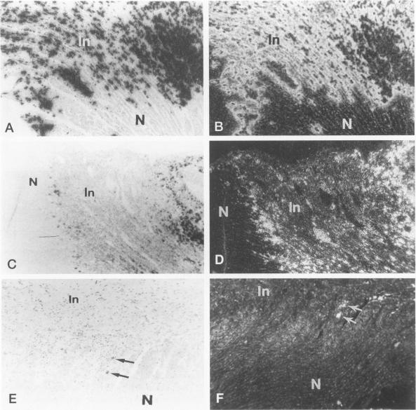
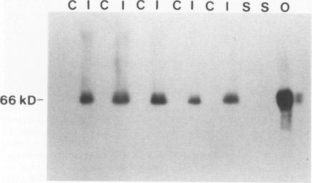
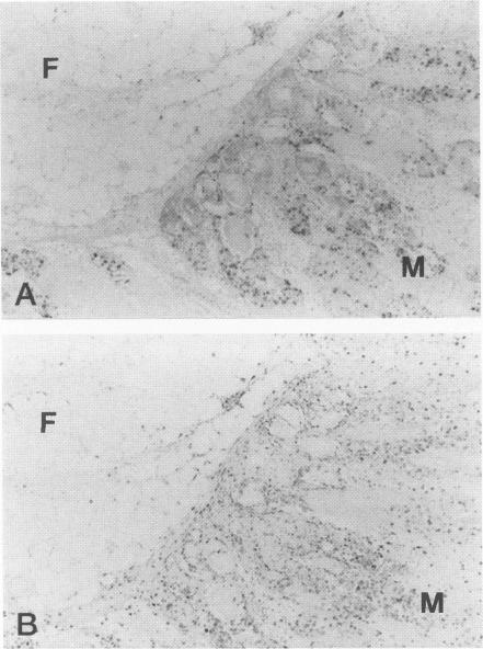
Osteopontin is a secreted glycoprotein implicated in a variety of functions, including cell adhesion and migration. Because these functions may be of general importance in the response of tissue to injury, we examined osteopontin expression after experimental cardiac injury and human myocardial infarction. Rat hearts were injured by transdiaphragmatic freeze-thaw and examined from 1 to 28 days after injury. Osteopontin was absent from normal myocardium by immunocytochemistry, Western blotting, and in situ hybridization. On days 1 and 2 after injury, osteopontin mRNA and protein were expressed at high levels by macrophages infiltrating necrotic myocardium. Double labeling with the macrophage marker ED1, however, demonstrated that only a subset of macrophages expressed osteopontin. Western blot analysis showed a single 66-kd band in injured myocardium that was absent from control tissue. Although macrophages remained abundant in the ensuing granulation response and scar tissue formation, the expression of osteopontin was diminished on day 4 and markedly downregulated at 1 and 4 weeks after injury, with only rare cells expressing the message or protein. In a human heart with an 8-day-old myocardial infarct, there was abundant expression of osteopontin mRNA and protein in macrophages within the necrotic and granulation tissue. Transient expression of osteopontin was also observed in a subset of macrophages infiltrating lung, skin, and skeletal muscle injured during the experiment, indicating the response was not limited to the heart. Thus, synthesis of osteopontin by macrophages appears to be a generalized response in the reaction to tissue injury. Although macrophages persist in these lesions, osteopontin is dramatically downregulated as healing proceeds. These results provide the first evidence that osteopontin may be important in healing after tissue injury, possibly in cellular adhesion, chemotaxis, and/or phagocytosis.

摘要

骨桥蛋白是一种分泌型糖蛋白,参与多种功能,包括细胞黏附和迁移。由于这些功能在组织对损伤的反应中可能具有普遍重要性,我们研究了实验性心脏损伤和人类心肌梗死后骨桥蛋白的表达情况。通过经膈冷冻-解冻法损伤大鼠心脏,并在损伤后1至28天进行检查。通过免疫细胞化学、蛋白质印迹法和原位杂交,正常心肌中未检测到骨桥蛋白。在损伤后第1天和第2天,浸润坏死心肌的巨噬细胞高水平表达骨桥蛋白mRNA和蛋白质。然而,用巨噬细胞标志物ED1进行双重标记显示,只有一部分巨噬细胞表达骨桥蛋白。蛋白质印迹分析显示,损伤心肌中有一条66-kd的条带,对照组织中则没有。尽管在随后的肉芽组织反应和瘢痕组织形成过程中巨噬细胞仍然大量存在,但骨桥蛋白的表达在第4天减少,在损伤后1周和4周明显下调,只有极少数细胞表达该信息或蛋白质。在一个有8天陈旧性心肌梗死的人心脏中,坏死和肉芽组织中的巨噬细胞大量表达骨桥蛋白mRNA和蛋白质。在实验过程中,浸润受伤肺、皮肤和骨骼肌的一部分巨噬细胞中也观察到骨桥蛋白的短暂表达,表明这种反应并不局限于心脏。因此,巨噬细胞合成骨桥蛋白似乎是对组织损伤反应中的一种普遍反应。尽管巨噬细胞持续存在于这些病变中,但随着愈合的进行,骨桥蛋白显著下调。这些结果首次证明骨桥蛋白可能在组织损伤后的愈合中起重要作用,可能参与细胞黏附、趋化性和/或吞噬作用。

https://cdn.ncbi.nlm.nih.gov/pmc/blobs/ba78/1887495/4c2f745df361/amjpathol00060-0220-a.jpg
https://cdn.ncbi.nlm.nih.gov/pmc/blobs/ba78/1887495/54eaab96312f/amjpathol00060-0216-a.jpg
https://cdn.ncbi.nlm.nih.gov/pmc/blobs/ba78/1887495/34ac07a96560/amjpathol00060-0217-a.jpg
https://cdn.ncbi.nlm.nih.gov/pmc/blobs/ba78/1887495/65f09f6cb56d/amjpathol00060-0218-a.jpg
https://cdn.ncbi.nlm.nih.gov/pmc/blobs/ba78/1887495/5b040a42aa4a/amjpathol00060-0218-b.jpg
https://cdn.ncbi.nlm.nih.gov/pmc/blobs/ba78/1887495/b8dfc316390f/amjpathol00060-0219-a.jpg
https://cdn.ncbi.nlm.nih.gov/pmc/blobs/ba78/1887495/bb2987b8ea05/amjpathol00060-0219-b.jpg
https://cdn.ncbi.nlm.nih.gov/pmc/blobs/ba78/1887495/4c2f745df361/amjpathol00060-0220-a.jpg

相似文献

1
Macrophages express osteopontin during repair of myocardial necrosis.巨噬细胞在心肌坏死修复过程中表达骨桥蛋白。
Am J Pathol. 1994 Dec;145(6):1450-62.
2
Spatially and temporally different expression of osteonectin and osteopontin in the infarct zone of experimentally induced myocardial infarction in rats.骨连接蛋白和骨桥蛋白在大鼠实验性心肌梗死梗死区的时空差异表达
Cardiovasc Pathol. 2003 Jul-Aug;12(4):186-94. doi: 10.1016/s1054-8807(03)00042-5.
3
Secretion of Osteopontin by macrophages and its accumulation at tissue surfaces during wound healing in mineralized tissues: a potential requirement for macrophage adhesion and phagocytosis.巨噬细胞分泌骨桥蛋白及其在矿化组织伤口愈合过程中在组织表面的积累:巨噬细胞黏附和吞噬作用的潜在需求。
Anat Rec. 1996 Jun;245(2):394-409. doi: 10.1002/(SICI)1097-0185(199606)245:2<394::AID-AR19>3.0.CO;2-K.
4
Temporal expression of osteopontin and CD44 in rat brains with experimental cryolesions.骨桥蛋白和CD44在实验性冷冻损伤大鼠脑内的时间表达
Brain Res. 2005 Apr 11;1041(1):95-101. doi: 10.1016/j.brainres.2005.02.019.
5
Expression of osteopontin in Kupffer cells and hepatic macrophages and Stellate cells in rat liver after carbon tetrachloride intoxication: a possible factor for macrophage migration into hepatic necrotic areas.四氯化碳中毒后大鼠肝脏中枯否细胞、肝巨噬细胞和星状细胞中骨桥蛋白的表达:巨噬细胞迁移至肝坏死区域的一个可能因素。
Biochem Biophys Res Commun. 1999 Mar 24;256(3):527-31. doi: 10.1006/bbrc.1999.0372.
6
Macrophage depletion impairs wound healing and increases left ventricular remodeling after myocardial injury in mice.巨噬细胞耗竭会损害小鼠伤口愈合,并加重心肌损伤后的左心室重塑。
Am J Pathol. 2007 Mar;170(3):818-29. doi: 10.2353/ajpath.2007.060547.
7
Increased expression of osteopontin in the heart tissue of Lewis rats with experimental autoimmune myocarditis.实验性自身免疫性心肌炎Lewis大鼠心脏组织中骨桥蛋白表达增加。
J Vet Med Sci. 2006 Apr;68(4):379-82. doi: 10.1292/jvms.68.379.
8
Muscle differentiation during repair of myocardial necrosis in rats via gene transfer with MyoD.通过MyoD基因转移修复大鼠心肌坏死过程中的肌肉分化
J Clin Invest. 1996 Nov 15;98(10):2209-17. doi: 10.1172/JCI119030.
9
Osteopontin expression is increased in the heritable cardiomyopathy of Syrian hamsters.骨桥蛋白的表达在叙利亚仓鼠的遗传性心肌病中增加。
Circulation. 1995 Aug 15;92(4):705-9. doi: 10.1161/01.cir.92.4.705.
10
Myocardial injury-induced fibroblast proliferation facilitates retroviral-mediated gene transfer to the rat heart in vivo.心肌损伤诱导的成纤维细胞增殖促进逆转录病毒介导的基因在大鼠心脏中的体内转移。
J Gene Med. 2000 Jan-Feb;2(1):2-10. doi: 10.1002/(SICI)1521-2254(200001/02)2:1<2::AID-JGM83>3.0.CO;2-O.

引用本文的文献

1
Osteopontin Expression and Its Role in Endometrial Cancer: A Systematic Review.骨桥蛋白的表达及其在子宫内膜癌中的作用:一项系统综述
Cancers (Basel). 2025 Jul 4;17(13):2245. doi: 10.3390/cancers17132245.
2
Post-myocardial Infarction Cardiac Remodeling: Multidimensional Mechanisms and Clinical Prospects of Stem Cell Therapy.心肌梗死后心脏重塑:干细胞治疗的多维机制与临床前景
Stem Cell Rev Rep. 2025 May 5. doi: 10.1007/s12015-025-10888-7.
3
Single-cell and spatial transcriptomics of the infarcted heart define the dynamic onset of the border zone in response to mechanical destabilization.

本文引用的文献

1
A re-examination of the molecular basis of cell movement.细胞运动分子基础的重新审视。
Immunol Cell Biol. 1993 Apr;71 ( Pt 2):131-9. doi: 10.1038/icb.1993.14.
2
Pattern of gene expression following rat tibial marrow ablation.大鼠胫骨骨髓消融后的基因表达模式。
J Bone Miner Res. 1993 Mar;8(3):379-88. doi: 10.1002/jbmr.5650080315.
3
Adhesion of metastatic, ras-transformed NIH 3T3 cells to osteopontin, fibronectin, and laminin.转移性、经ras基因转化的NIH 3T3细胞与骨桥蛋白、纤连蛋白和层粘连蛋白的黏附作用。
梗死心脏的单细胞和空间转录组学确定了对机械失稳作出反应的边缘区的动态起始过程。
Nat Cardiovasc Res. 2022 Nov;1(11):1039-1055. doi: 10.1038/s44161-022-00160-3. Epub 2022 Nov 17.
4
Don't go breakin' my heart: cardioprotective alterations to the mechanical and structural properties of reperfused myocardium during post-infarction inflammation.别伤我的心:心肌梗死后炎症期间再灌注心肌机械和结构特性的心脏保护改变
Biophys Rev. 2023 Jun 10;15(3):329-353. doi: 10.1007/s12551-023-01068-3. eCollection 2023 Jun.
5
Extracellular matrix stiffness-The central cue for skin fibrosis.细胞外基质硬度——皮肤纤维化的核心线索。
Front Mol Biosci. 2023 Mar 8;10:1132353. doi: 10.3389/fmolb.2023.1132353. eCollection 2023.
6
Integrated analysis of potential gene crosstalk between non-alcoholic fatty liver disease and diabetic nephropathy.非酒精性脂肪性肝病与糖尿病肾病之间潜在基因相互作用的综合分析。
Front Endocrinol (Lausanne). 2022 Oct 25;13:1032814. doi: 10.3389/fendo.2022.1032814. eCollection 2022.
7
The Patent Ductus Arteriosus in Extremely Preterm Neonates Is More than a Hemodynamic Challenge: New Molecular Insights.动脉导管未闭在极早产儿中不仅仅是一个血流动力学挑战:新的分子见解。
Biomolecules. 2022 Aug 25;12(9):1179. doi: 10.3390/biom12091179.
8
Macrophages in myocardial infarction.心肌梗死中的巨噬细胞。
Am J Physiol Cell Physiol. 2022 Oct 1;323(4):C1304-C1324. doi: 10.1152/ajpcell.00230.2022. Epub 2022 Sep 12.
9
Clinical and Molecular Implications of Osteopontin in Heart Failure.骨桥蛋白在心力衰竭中的临床及分子学意义
Curr Issues Mol Biol. 2022 Aug 11;44(8):3573-3597. doi: 10.3390/cimb44080245.
10
The role of endogenous Smad7 in regulating macrophage phenotype following myocardial infarction.内源性 Smad7 在心肌梗死后调节巨噬细胞表型中的作用。
FASEB J. 2022 Jul;36(7):e22400. doi: 10.1096/fj.202101956RR.
Cancer Res. 1993 Feb 1;53(3):701-6.
4
Osteopontin overexpression is associated with arterial smooth muscle cell proliferation in vitro.骨桥蛋白过表达与体外动脉平滑肌细胞增殖有关。
Arterioscler Thromb. 1993 Jan;13(1):120-5. doi: 10.1161/01.atv.13.1.120.
5
Osteopontin is elevated during neointima formation in rat arteries and is a novel component of human atherosclerotic plaques.骨桥蛋白在大鼠动脉新生内膜形成过程中升高,是人类动脉粥样硬化斑块的一种新成分。
J Clin Invest. 1993 Oct;92(4):1686-96. doi: 10.1172/JCI116755.
6
Mechanism of fibronectin enhancement of group B streptococcal phagocytosis by human neutrophils and culture-derived macrophages.纤连蛋白增强人中性粒细胞和培养来源巨噬细胞对B族链球菌吞噬作用的机制。
Infect Immun. 1993 Jun;61(6):2334-9. doi: 10.1128/iai.61.6.2334-2339.1993.
7
Expression of osteopontin messenger RNA by macrophages in atherosclerotic plaques. A possible association with calcification.动脉粥样硬化斑块中巨噬细胞骨桥蛋白信使核糖核酸的表达。与钙化的可能关联。
Am J Pathol. 1993 Oct;143(4):1003-8.
8
Osteopontin expression in angiotensin II-induced tubulointerstitial nephritis.骨桥蛋白在血管紧张素II诱导的肾小管间质性肾炎中的表达
Kidney Int. 1994 Feb;45(2):515-24. doi: 10.1038/ki.1994.67.
9
Proliferation in primary and restenotic coronary atherectomy tissue. Implications for antiproliferative therapy.原发性及再狭窄冠状动脉旋切组织中的增殖。抗增殖治疗的意义。
Circ Res. 1993 Aug;73(2):223-31. doi: 10.1161/01.res.73.2.223.
10
Isolation and characterization of two sialoproteins present only in bone calcified matrix.仅存在于骨钙化基质中的两种唾液蛋白的分离与鉴定。
Biochem J. 1985 Dec 15;232(3):715-24. doi: 10.1042/bj2320715.