Brouwer M, Hoexum-Brouwer T, Cashon R E
Duke University School of the Environment, Marine Laboratory/Marine Biomedical Center, Beaufort, NC 28516.
Biochem J. 1993 Aug 15;294 ( Pt 1)(Pt 1):219-25. doi: 10.1042/bj2940219.
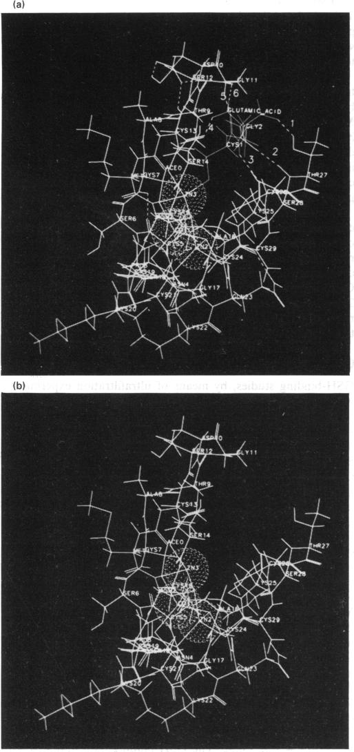
Glutathione (GSH) has been found to form a complex with both vertebrate and invertebrate copper-metallothionein (CuMT) [Freedman, Ciriolo and Peisach (1989) J. Biol. Chem. 264, 5598-5605; Brouwer and Brouwer-Hoexum (1991) Arch. Biochem. Biophys. 290, 207-213]. In this paper we report on the interaction of GSH with CdZnMT-I and CdZnMT-II from rabbit liver and with CdMT-I from Blue crab hepatopancreas. Ultrafiltration experiments showed that all three MTs combined with GSH. The measured binding data for the three MTs could be described by a single binding isotherm. The GSH/MT stoichiometry was 1.4 +/- 0.3 and Kdiss. = 14 +/- 6 microM. Partially Zn-depleted MT does not significantly bind GSH, indicating that the GSH-binding site is located on MT's Zn-containing N-terminal domain. The putative GSH-binding site on rabbit liver MT was investigated using molecular-graphics analysis. A cleft on the MT's N-terminal domain, which has the labile Zn-2 at its base, could easily accommodate GSH. Cysteine-ligand exchange between the terminal (non-bridging) Cys-26, bound to Zn-2, and the cysteine in GSH is stereochemically possible. Based on these considerations a model of MT-GSH was built in which GSH's cysteine replaces Cys-26 as a terminal Zn-2 ligand. This complex was energy-minimized by molecular-mechanics calculations, taking into account computed partial electrostatic charges on all atoms, including Cd and Zn. These calculations showed that the MT-GSH complex was thermodynamically more stable than MT, due to favourable non-bonded, electrostatic and van der Waals interactions. Six hydrogen bonds can form between GSH and MT. The average pairwise root-mean-square deviations (RMSD) of the metals in energy-minimized MT and MT-GSH, compared with the metals in the crystal structure, were 0.0087 +/- 0.0028 nm (0.087 +/- 0.028 A) and 0.0168 +/- 0.0087 nm (0.168 +/- 0.087 A) respectively. The RMSD values for the polypeptide-backbone alpha carbons were 0.0136 +/- 0.0060 nm (0.136 +/- 0.060 A) and 0.0491 +/- 0.0380 nm (0.491 +/- 0.380 A) respectively. No other docking sites for GSH were found. The energy-minimized structure of an MT-2-mercaptoethanol complex was somewhat less stable than the native MT domain, attesting to the specificity of the MT-GSH interaction. The possible physiological significance of the MT-GSH interaction is discussed.
已发现谷胱甘肽(GSH)可与脊椎动物和无脊椎动物的铜金属硫蛋白(CuMT)形成复合物[弗里德曼、西里奥洛和佩萨奇(1989年)《生物化学杂志》264卷,5598 - 5605页;布劳威尔和布劳威尔 - 霍克苏姆(1991年)《生物化学与生物物理学报》290卷,207 - 213页]。在本文中,我们报告了GSH与兔肝中的CdZnMT - I和CdZnMT - II以及蓝蟹肝胰腺中的CdMT - I之间的相互作用。超滤实验表明,所有三种金属硫蛋白均与GSH结合。三种金属硫蛋白的实测结合数据可用单一结合等温线描述。GSH/金属硫蛋白化学计量比为1.4±0.3,解离常数Kdiss. = 14±6微摩尔。部分锌缺失的金属硫蛋白不与GSH显著结合,表明GSH结合位点位于金属硫蛋白含锌的N端结构域。利用分子图形分析研究了兔肝金属硫蛋白上假定的GSH结合位点。金属硫蛋白N端结构域上的一个裂隙,其底部有不稳定的Zn - 2,能够轻松容纳GSH。与Zn - 2结合的末端(非桥连)半胱氨酸 - 26与GSH中的半胱氨酸之间的半胱氨酸 - 配体交换在立体化学上是可能的。基于这些考虑,构建了一个金属硫蛋白 - GSH模型,其中GSH的半胱氨酸取代半胱氨酸 - 26作为末端Zn - 2配体。通过分子力学计算对该复合物进行了能量最小化处理,同时考虑了包括镉和锌在内的所有原子的计算部分静电荷。这些计算表明,由于有利的非键合、静电和范德华相互作用,金属硫蛋白 - GSH复合物在热力学上比金属硫蛋白更稳定。GSH与金属硫蛋白之间可形成六个氢键。与晶体结构中的金属相比,能量最小化后的金属硫蛋白和金属硫蛋白 - GSH中金属的平均成对均方根偏差(RMSD)分别为0.0087±0.0028纳米(0.087±0.028埃)和0.0168±0.0087纳米(0.168±0.087埃)。多肽主链α碳原子的RMSD值分别为0.0136±0.0060纳米(0.136±0.060埃)和0.0491±0.0380纳米(0.491±0.380埃)。未发现其他GSH对接位点。金属硫蛋白 - 2 - 巯基乙醇复合物的能量最小化结构比天然金属硫蛋白结构域稍不稳定,这证明了金属硫蛋白 - GSH相互作用的特异性。讨论了金属硫蛋白 - GSH相互作用可能的生理意义。