• 文献检索
  • 文档翻译
  • 深度研究
  • 学术资讯
  • Suppr Zotero 插件Zotero 插件
  • 邀请有礼
  • 套餐&价格
  • 历史记录
应用&插件
Suppr Zotero 插件Zotero 插件浏览器插件Mac 客户端Windows 客户端微信小程序
定价
高级版会员购买积分包购买API积分包
服务
文献检索文档翻译深度研究API 文档MCP 服务
关于我们
关于 Suppr公司介绍联系我们用户协议隐私条款
关注我们

Suppr 超能文献

核心技术专利:CN118964589B侵权必究
粤ICP备2023148730 号-1Suppr @ 2026

文献检索

告别复杂PubMed语法,用中文像聊天一样搜索,搜遍4000万医学文献。AI智能推荐,让科研检索更轻松。

立即免费搜索

文件翻译

保留排版,准确专业,支持PDF/Word/PPT等文件格式,支持 12+语言互译。

免费翻译文档

深度研究

AI帮你快速写综述,25分钟生成高质量综述,智能提取关键信息,辅助科研写作。

立即免费体验

通过X射线衍射、拉曼光谱和分子建模研究晶体和溶液中d(GGGATCCC)2的构象。

Conformation of d(GGGATCCC)2 in crystals and in solution studied by X-ray diffraction, Raman spectroscopy and molecular modelling.

作者信息

Fabian H, Hölzer W, Heinemann U, Sklenar H, Welfle H

机构信息

Max-Delbrück-Center for Molecular Medicine, Berlin-Buch, Germany.

出版信息

Nucleic Acids Res. 1993 Feb 11;21(3):569-76. doi: 10.1093/nar/21.3.569.

DOI:10.1093/nar/21.3.569
PMID:8441669
原文链接:https://pmc.ncbi.nlm.nih.gov/articles/PMC309154/
Abstract

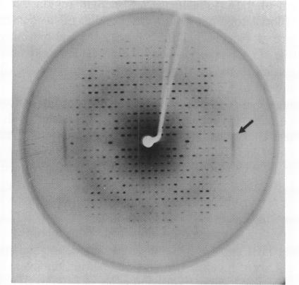
In the crystal, d(GGGATCCC)2 forms an A-DNA double helix as known from a single crystal X-ray diffraction study. Accordingly, in the Raman spectra of crystals the A-family marker bands at 664, 705, 807 and 1101 cm-1 and the spectral characteristics in the region 1200 to 1500 cm-1 clearly demonstrate the A-form as the dominant conformation. Bands at 691, 850, and 1080 cm-1, however, indicate that a minor fraction of the octamer molecules in the crystal is in an unusual, still not unequivocally identified conformation possibly belonging to the B-family. In solution, the octamer is in B-like conformation as shown by the presence of B-DNA Raman marker bands at 685, 837, 1094 and 1421 cm-1. Molecular modelling techniques lead to three structures with slightly different B-form geometries as the lowest energies models when a sigmoidal dielectric function with the bulk dielectric constant epsilon = 78 and the value q = -0.5e for the effective phosphate charges was used in the calculations. An A-form structure bearing a strong resemblance to the experimentally determined crystal structure becomes the lowest energy model structure when the electrostatic parameters are changed to epsilon = 30 and q = -0.25e, respectively.

摘要

在晶体中,d(GGGATCCC)2形成了一种A-DNA双螺旋结构,这是通过单晶X射线衍射研究得知的。因此,在晶体的拉曼光谱中,664、705、807和1101 cm-1处的A家族标记带以及1200至1500 cm-1区域的光谱特征清楚地表明A构象是主要构象。然而,691、850和1080 cm-1处的谱带表明,晶体中八聚体分子的一小部分处于一种异常的、尚未明确鉴定的构象,可能属于B家族。在溶液中,八聚体呈类B构象,685、837、1094和1421 cm-1处的B-DNA拉曼标记带的出现表明了这一点。当在计算中使用具有体介电常数ε = 78和有效磷酸电荷值q = -0.5e的S形介电函数时,分子建模技术得出了三种具有略有不同B形几何结构的结构作为最低能量模型。当静电参数分别变为ε = 30和q = -0.25e时,一种与实验确定的晶体结构非常相似的A形结构成为最低能量模型结构。

https://cdn.ncbi.nlm.nih.gov/pmc/blobs/33b0/309154/a06aee4ae3db/nar00052-0209-a.jpg

相似文献

1
Conformation of d(GGGATCCC)2 in crystals and in solution studied by X-ray diffraction, Raman spectroscopy and molecular modelling.通过X射线衍射、拉曼光谱和分子建模研究晶体和溶液中d(GGGATCCC)2的构象。
Nucleic Acids Res. 1993 Feb 11;21(3):569-76. doi: 10.1093/nar/21.3.569.
2
Raman spectra of single crystals of r(GCG)d(CGC) and d(CCCCGGGG) as models for A DNA, their structure transitions in aqueous solution, and comparison with double-helical poly(dG).poly(dC).以r(GCG)d(CGC)和d(CCCCGGGG)作为A-DNA模型的单晶拉曼光谱、它们在水溶液中的结构转变以及与双螺旋聚(dG).聚(dC)的比较
Biochemistry. 1986 Jan 14;25(1):41-50. doi: 10.1021/bi00349a007.
3
The infrared and Raman spectra of the duplex of d(GGTATACC) in the crystal show bands due to both the A-form and the B-form of DNA.晶体中d(GGTATACC)双链体的红外光谱和拉曼光谱显示出归因于DNA的A形式和B形式的谱带。
J Biomol Struct Dyn. 1990 Oct;8(2):295-302. doi: 10.1080/07391102.1990.10507806.
4
Crystal and solution structures of the B-DNA dodecamer d(CGCAAATTTGCG) probed by Raman spectroscopy: heterogeneity in the crystal structure does not persist in the solution structure.通过拉曼光谱探测的B-DNA十二聚体d(CGCAAATTTGCG)的晶体结构和溶液结构:晶体结构中的异质性在溶液结构中并不持续存在。
Biochemistry. 1988 Feb 9;27(3):931-8. doi: 10.1021/bi00403a014.
5
Evidence for crystal environment dominating base sequence effects on DNA conformation: crystal structures of the orthorhombic and hexagonal polymorphs of the A-DNA decamer d(GCGGGCCCGC) and comparison with their isomorphous crystal structures.晶体环境主导碱基序列对DNA构象影响的证据:A-DNA十聚体d(GCGGGCCCGC)正交晶型和六方晶型的晶体结构及其同晶型晶体结构的比较
Biochemistry. 1993 Oct 26;32(42):11458-68. doi: 10.1021/bi00093a025.
6
Orthorhombic crystal structure of the A-DNA octamer d(GTACGTAC). Comparison with the tetragonal structure.
Eur J Biochem. 1993 Apr 15;213(2):673-82. doi: 10.1111/j.1432-1033.1993.tb17807.x.
7
Demonstration of Z-d(5BrCGAT5BrCG) and B-d(CGCGATCGCG) form crystal structures in DNA-cobalt hexammine complexes by Kr 647.1 nm excitation of Raman spectra.通过氪647.1纳米激发拉曼光谱法,证明DNA-六氨合钴配合物中Z-d(5BrCGAT5BrCG)和B-d(CGCGATCGCG)形成晶体结构。
Nucleic Acids Res. 1993 Mar 25;21(6):1433-8. doi: 10.1093/nar/21.6.1433.
8
Structure of the B-DNA oligomers d(CGCTAGCG) and d(CGCTCTAGAGCG) in new crystal forms.新型晶体形式下B-DNA寡聚物d(CGCTAGCG)和d(CGCTCTAGAGCG)的结构
Biochemistry. 1996 Sep 10;35(36):11589-95. doi: 10.1021/bi961048l.
9
Effect of the G.T mismatch on backbone and sugar conformations of Z-DNA and B-DNA: analysis by Raman spectroscopy of crystal and solution structures of d(CGCGTG) and d(CGCGCG).G.T错配对Z-DNA和B-DNA骨架及糖构象的影响:通过拉曼光谱分析d(CGCGTG)和d(CGCGCG)的晶体及溶液结构
Biochemistry. 1989 Jan 10;28(1):304-10. doi: 10.1021/bi00427a041.
10
Conformations in crystals and solutions of d(CACGTG), d(CCGCGG) and d(GGCGCC) studied by vibrational spectroscopy.通过振动光谱学研究d(CACGTG)、d(CCGCGG)和d(GGCGCC)在晶体和溶液中的构象。
Nucleic Acids Res. 1989 Aug 25;17(16):6669-80. doi: 10.1093/nar/17.16.6669.

引用本文的文献

1
The interactions of monomeric acridines and unsymmetrical bisacridines (UAs) with DNA duplexes: an insight provided by NMR and MD studies.单体吖啶和不对称双吖啶(UAs)与 DNA 双链体的相互作用:NMR 和 MD 研究提供的深入了解。
Sci Rep. 2023 Mar 1;13(1):3431. doi: 10.1038/s41598-023-30587-y.
2
Comparison of the solution and crystal conformations of (G + C)-rich fragments of DNA.DNA中富含(G + C)片段的溶液构象与晶体构象的比较。
Biophys J. 1996 Sep;71(3):1530-8. doi: 10.1016/S0006-3495(96)79355-4.

本文引用的文献

1
Changes in raman vibrational bands of calf thymus DNA during the B-to-A transition.小牛胸腺DNA从B型向A型转变过程中拉曼振动带的变化。
Biopolymers. 1982 Mar;21(3):499-512. doi: 10.1002/bip.360210303.
2
Sequence-dependent conformation of an A-DNA double helix. The crystal structure of the octamer d(G-G-T-A-T-A-C-C).A-DNA双螺旋的序列依赖性构象。八聚体d(G-G-T-A-T-A-C-C)的晶体结构。
J Mol Biol. 1983 May 15;166(2):183-201. doi: 10.1016/s0022-2836(83)80005-9.
3
Raman spectra of single crystals of r(GCG)d(CGC) and d(CCCCGGGG) as models for A DNA, their structure transitions in aqueous solution, and comparison with double-helical poly(dG).poly(dC).
以r(GCG)d(CGC)和d(CCCCGGGG)作为A-DNA模型的单晶拉曼光谱、它们在水溶液中的结构转变以及与双螺旋聚(dG).聚(dC)的比较
Biochemistry. 1986 Jan 14;25(1):41-50. doi: 10.1021/bi00349a007.
4
Characteristics and variations of B-type DNA conformations in solution: a quantitative analysis of Raman band intensities of eight DNAs.溶液中B型DNA构象的特征与变异:八种DNA拉曼谱带强度的定量分析
Biochemistry. 1986 May 6;25(9):2664-71. doi: 10.1021/bi00357a056.
5
Crystal structure analysis of an A-DNA fragment at 1.8 A resolution: d(GCCCGGGC).分辨率为1.8埃的A-DNA片段d(GCCCGGGC)的晶体结构分析
Nucleic Acids Res. 1987 Nov 25;15(22):9531-50. doi: 10.1093/nar/15.22.9531.
6
The crystal structure of d(CCCCGGGG): a new A-form variant with an extended backbone conformation.d(CCCCGGGG)的晶体结构:一种具有延伸主链构象的新型A 型变体。
J Biomol Struct Dyn. 1987 Oct;5(2):199-217. doi: 10.1080/07391102.1987.10506390.
7
A general approach to the optimization of the conformation of ring molecules with an application to valinomycin.一种优化环状分子构象的通用方法及其在缬氨霉素中的应用。
J Biomol Struct Dyn. 1986 Dec;4(3):443-62. doi: 10.1080/07391102.1986.10506361.
8
The flexibility of the nucleic acids: (II). The calculation of internal energy and applications to mononucleotide repeat DNA.核酸的柔韧性:(II)。单核苷酸重复DNA的内能计算及应用
J Biomol Struct Dyn. 1986 Apr;3(5):989-1014. doi: 10.1080/07391102.1986.10508478.
9
The flexibility of the nucleic acids: (I). "SIR", a novel approach to the variation of polymer geometry in constrained systems.核酸的柔韧性:(I). “SIR”,一种在受限系统中改变聚合物几何形状的新方法。
J Biomol Struct Dyn. 1986 Apr;3(5):967-87. doi: 10.1080/07391102.1986.10508477.
10
Three-dimensional structure of d(GGGATCCC) in the crystalline state.
Nucleic Acids Res. 1988 Aug 25;16(16):7799-816. doi: 10.1093/nar/16.16.7799.