Adamson I Y, Bakowska J, Bowden D H
Department of Pathology, University of Manitoba, Winnipeg, Canada.
Am J Pathol. 1993 Apr;142(4):1209-16.
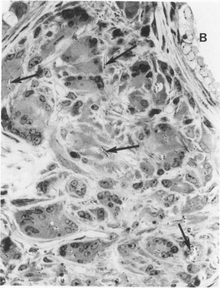
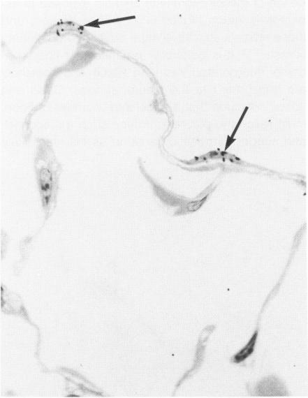
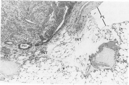
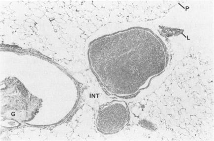
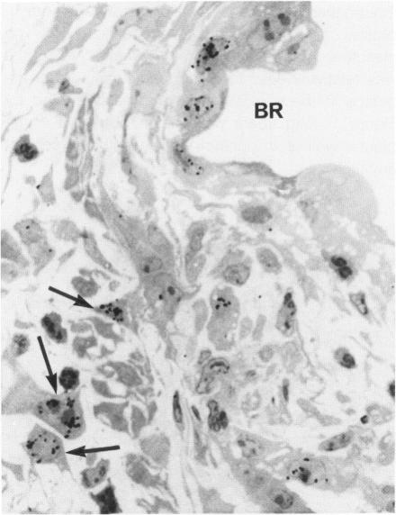
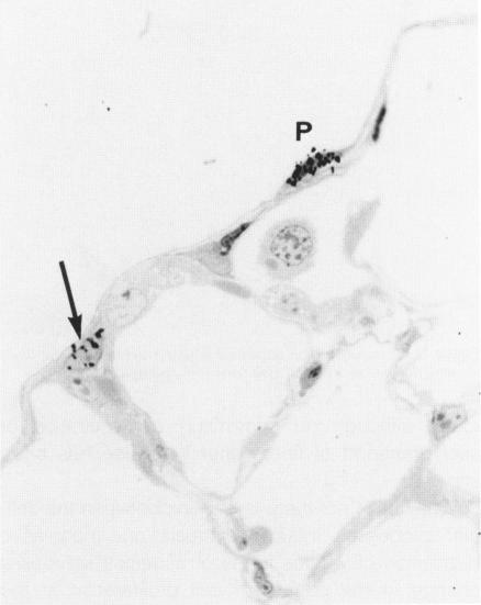
The relationship of asbestos deposition in the lung to subsequent cell proliferation at the pleural surface is not clear. The present study examines DNA synthesis by various pulmonary cells, particularly those at the pleura after intratracheal injection of 0.1 mg crocidolite to mice using: 1) long fibers (> 20 mu), which are deposited in bronchiolar regions and induce fibrosis; 2) short fibers (< 1 mu), which reach alveoli but do not induce fibrosis. Mice also received 2 microCi/g tritiated thymidine 1 hour before death at intervals to 16 weeks. Short fibers induced only a small increase in labeling of bronchiolar epithelial and interstitial cells, which subsided by 5 days, when a small increase in labeled mesothelial and subpleural cells was seen. In contrast, long fibers damaged the bronchiolar epithelium and became incorporated into connective tissue. During regeneration, 12% of cells were labeled at 3 days and labeling was greater than controls to 4 weeks. Increased peribronchiolar labeling of fibroblasts and interstitial macrophages was seen around long fibers, and increased DNA synthesis by mesothelial and subpleural cells was found. Up to 2% of mesothelial cells were labeled 1 week after long fibers compared to near zero in controls. No long fibers were found at the pleura. Activation of interstitial macrophages in response to long crocidolite fibers is associated with fibroblast proliferation. It is now suggested that mesothelial cells may also be stimulated by cytokines from activated interstitial macrophages that diffuse across the interstitium, without requiring actual fiber translocation to the pleura.
肺中石棉沉积与随后胸膜表面细胞增殖之间的关系尚不清楚。本研究通过气管内给小鼠注射0.1mg青石棉,使用以下方法检测各种肺细胞,特别是胸膜处细胞的DNA合成:1)长纤维(>20μm),其沉积在细支气管区域并诱导纤维化;2)短纤维(<1μm),其到达肺泡但不诱导纤维化。小鼠在处死前1小时每隔一定时间接受2μCi/g的氚标记胸腺嘧啶核苷,最长至16周。短纤维仅使细支气管上皮细胞和间质细胞的标记略有增加,5天后这种增加就消退了,此时可见间皮细胞和胸膜下细胞的标记略有增加。相比之下,长纤维损伤了细支气管上皮并融入结缔组织。在再生过程中,3天时12%的细胞被标记,并且标记在4周内均高于对照组。在长纤维周围可见成纤维细胞和间质巨噬细胞的细支气管周围标记增加,并且发现间皮细胞和胸膜下细胞的DNA合成增加。长纤维处理1周后,高达2%的间皮细胞被标记,而对照组接近零。在胸膜处未发现长纤维。对长青石棉纤维的反应中,间质巨噬细胞的激活与成纤维细胞增殖有关。现在有人提出,间皮细胞也可能受到来自活化间质巨噬细胞的细胞因子的刺激,这些细胞因子通过间质扩散,而不需要纤维实际转移到胸膜。