Huynh H K, Dorovini-Zis K
Department of Pathology, Vancouver General Hospital, British Columbia, Canada.
Am J Pathol. 1993 Apr;142(4):1265-78.
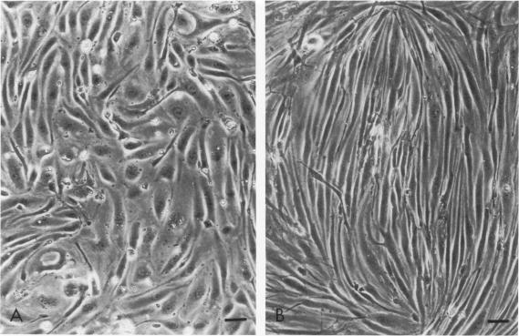
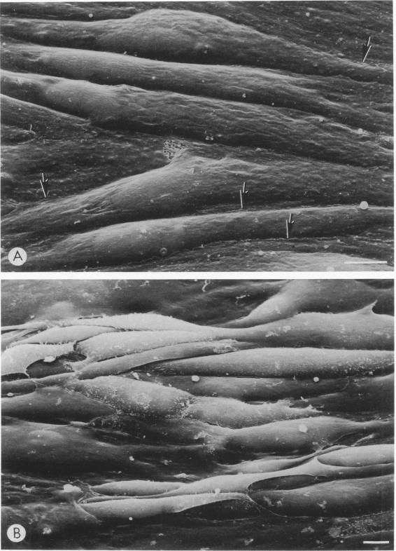
Primary cultures of human brain microvessel endothelial cells were used to study the effects of human recombinant interferon-gamma (IFN-gamma) on cerebral endothelium in vitro. Incubation of monolayers with various concentrations of IFN-gamma (10 to 200 U/ml) for 12 to 96 hours induced surface expression of class II major histocompatibility complex (Ia) antigen in a time- and concentration-dependent manner. In immunogold-stained cultures, labeling was observed as early as 12 hours, was maximal after 48 hours, and persisted at plateau levels in the continuous presence of the cytokine. Expression was blocked by coincubation with anti-IFN-gamma antibody and was reversed 4 days following removal of IFN-gamma from the culture media. Endothelial cells treated with IFN-gamma for 3 to 4 days became spindle-shaped, extensively overlapped, and frequently formed cellular whorls. These changes did not occur in the presence of anti-IFN-gamma antibody and reversed upon removal of IFN-gamma from the media. The morphological alterations were associated with increased permeability of confluent monolayers to macromolecules as compared with untreated cultures. The results of these studies indicate that human brain microvessel endothelial cells respond to in vitro cytokine stimulation by undergoing profound morphological, functional, and permeability changes. We conclude that cerebral endothelium may play an important role in the initiation and regulation of lymphocyte traffic across the blood-brain barrier in inflammatory disorders of the human central nervous system.
用人脑微血管内皮细胞的原代培养物来研究重组人γ干扰素(IFN-γ)对体外脑内皮细胞的影响。用不同浓度的IFN-γ(10至200 U/ml)处理单层细胞12至96小时,以时间和浓度依赖性方式诱导II类主要组织相容性复合体(Ia)抗原的表面表达。在免疫金染色的培养物中,最早在12小时观察到标记,48小时后达到最大值,并且在细胞因子持续存在的情况下维持在稳定水平。与抗IFN-γ抗体共同孵育可阻断表达,从培养基中去除IFN-γ 4天后表达恢复。用IFN-γ处理3至4天的内皮细胞变成纺锤形,广泛重叠,并经常形成细胞漩涡。在存在抗IFN-γ抗体的情况下不会发生这些变化,从培养基中去除IFN-γ后这些变化会逆转。与未处理的培养物相比,形态学改变与汇合单层对大分子的通透性增加有关。这些研究结果表明,人脑微血管内皮细胞通过经历深刻的形态、功能和通透性变化来响应体外细胞因子刺激。我们得出结论,在人类中枢神经系统炎症性疾病中,脑内皮细胞可能在淋巴细胞穿越血脑屏障的起始和调节中起重要作用。