• 文献检索
  • 文档翻译
  • 深度研究
  • 学术资讯
  • Suppr Zotero 插件Zotero 插件
  • 邀请有礼
  • 套餐&价格
  • 历史记录
应用&插件
Suppr Zotero 插件Zotero 插件浏览器插件Mac 客户端Windows 客户端微信小程序
定价
高级版会员购买积分包购买API积分包
服务
文献检索文档翻译深度研究API 文档MCP 服务
关于我们
关于 Suppr公司介绍联系我们用户协议隐私条款
关注我们

Suppr 超能文献

核心技术专利:CN118964589B侵权必究
粤ICP备2023148730 号-1Suppr @ 2026

文献检索

告别复杂PubMed语法,用中文像聊天一样搜索,搜遍4000万医学文献。AI智能推荐,让科研检索更轻松。

立即免费搜索

文件翻译

保留排版,准确专业,支持PDF/Word/PPT等文件格式,支持 12+语言互译。

免费翻译文档

深度研究

AI帮你快速写综述,25分钟生成高质量综述,智能提取关键信息,辅助科研写作。

立即免费体验

血管紧张素II 2型受体介导程序性细胞死亡。

Angiotensin II type 2 receptor mediates programmed cell death.

作者信息

Yamada T, Horiuchi M, Dzau V J

机构信息

Falk Cardiovascular Research Center, Stanford University School of Medicine, CA 94305-5246, USA.

出版信息

Proc Natl Acad Sci U S A. 1996 Jan 9;93(1):156-60. doi: 10.1073/pnas.93.1.156.

DOI:10.1073/pnas.93.1.156
PMID:8552595
原文链接:https://pmc.ncbi.nlm.nih.gov/articles/PMC40197/
Abstract

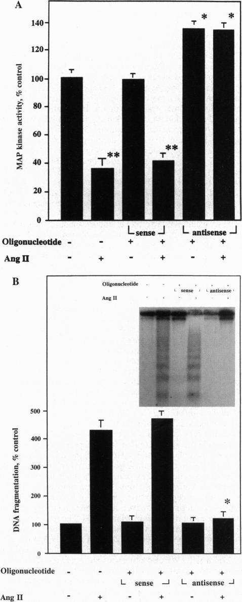
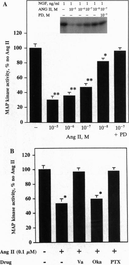
The function of the recently discovered angiotensin II type 2 (AT2) receptor remains elusive. This receptor is expressed abundantly in fetus, but scantily in adult tissues except brain, adrenal medulla, and atretic ovary. In this study, we demonstrated that this receptor mediates programmed cell death (apoptosis). We observed this effect in PC12W cells (rat pheochromocytoma cell line) and R3T3 cells (mouse fibroblast cell line), which express abundant AT2 receptor but not AT1 receptor. The cellular mechanism appears to involve the dephosphorylation of mitogen-activated protein kinase (MAP kinase). Vanadate, a protein-tyrosine-phosphatase inhibitor, attenuated the dephosphorylation of MAP kinases by the AT2 receptor and restored the apoptotic changes. Antisense oligonucleotide to MAP kinase phosphatase 1 inhibited the AT2 receptor-mediated MAP kinase dephosphorylation and blocked the AT2 receptor-mediated apoptosis. These results suggest that protein-tyrosine-phosphatase, including MAP kinase phosphatase 1 activated by the AT2 receptor, is involved in apoptosis. We hypothesize that this apoptotic function of the AT2 receptor may play an important role in developmental biology and pathophysiology.

摘要

最近发现的血管紧张素II 2型(AT2)受体的功能仍不清楚。该受体在胎儿中大量表达,但在成体组织中表达较少,除了脑、肾上腺髓质和闭锁卵巢。在本研究中,我们证明该受体介导程序性细胞死亡(凋亡)。我们在PC12W细胞(大鼠嗜铬细胞瘤细胞系)和R3T3细胞(小鼠成纤维细胞系)中观察到这种效应,这两种细胞系表达丰富的AT2受体但不表达AT1受体。细胞机制似乎涉及丝裂原活化蛋白激酶(MAP激酶)的去磷酸化。钒酸盐是一种蛋白酪氨酸磷酸酶抑制剂,它减弱了AT2受体介导的MAP激酶去磷酸化并恢复了凋亡变化。针对MAP激酶磷酸酶1的反义寡核苷酸抑制了AT2受体介导的MAP激酶去磷酸化并阻断了AT2受体介导的凋亡。这些结果表明,包括由AT2受体激活的MAP激酶磷酸酶1在内的蛋白酪氨酸磷酸酶参与了凋亡。我们推测AT2受体的这种凋亡功能可能在发育生物学和病理生理学中起重要作用。

https://cdn.ncbi.nlm.nih.gov/pmc/blobs/dcc8/40197/320d36fe2180/pnas01505-0170-b.jpg
https://cdn.ncbi.nlm.nih.gov/pmc/blobs/dcc8/40197/4fc029e8f9e9/pnas01505-0167-a.jpg
https://cdn.ncbi.nlm.nih.gov/pmc/blobs/dcc8/40197/7d782bfc8fc2/pnas01505-0168-a.jpg
https://cdn.ncbi.nlm.nih.gov/pmc/blobs/dcc8/40197/e24e3c2caaea/pnas01505-0169-a.jpg
https://cdn.ncbi.nlm.nih.gov/pmc/blobs/dcc8/40197/a51d1f840ea1/pnas01505-0169-b.jpg
https://cdn.ncbi.nlm.nih.gov/pmc/blobs/dcc8/40197/1578e77f4982/pnas01505-0170-a.jpg
https://cdn.ncbi.nlm.nih.gov/pmc/blobs/dcc8/40197/320d36fe2180/pnas01505-0170-b.jpg

相似文献

1
Angiotensin II type 2 receptor mediates programmed cell death.血管紧张素II 2型受体介导程序性细胞死亡。
Proc Natl Acad Sci U S A. 1996 Jan 9;93(1):156-60. doi: 10.1073/pnas.93.1.156.
2
Angiotensin type 2 receptor dephosphorylates Bcl-2 by activating mitogen-activated protein kinase phosphatase-1 and induces apoptosis.血管紧张素2型受体通过激活丝裂原活化蛋白激酶磷酸酶-1使Bcl-2去磷酸化并诱导细胞凋亡。
J Biol Chem. 1997 Jul 25;272(30):19022-6. doi: 10.1074/jbc.272.30.19022.
3
Angiotensin II type 2 receptors mediate inhibition of mitogen-activated protein kinase cascade and functional activation of SHP-1 tyrosine phosphatase.血管紧张素II 2型受体介导对丝裂原活化蛋白激酶级联反应的抑制及SHP-1酪氨酸磷酸酶的功能激活。
Biochem J. 1997 Jul 15;325 ( Pt 2)(Pt 2):449-54. doi: 10.1042/bj3250449.
4
Role of AT1 and AT2 receptors in regulation of MAPKs and MKP-1 by ANG II in adult cardiac myocytes.AT1和AT2受体在成年心肌细胞中血管紧张素II对丝裂原活化蛋白激酶和丝裂原活化蛋白激酶磷酸酶-1的调节作用。
Am J Physiol. 1998 Sep;275(3):H906-16. doi: 10.1152/ajpheart.1998.275.3.H906.
5
Inactivation of p42 MAP kinase by protein phosphatase 2A and a protein tyrosine phosphatase, but not CL100, in various cell lines.在多种细胞系中,蛋白磷酸酶2A和一种蛋白酪氨酸磷酸酶可使p42丝裂原活化蛋白激酶失活,但CL100不能。
Curr Biol. 1995 Mar 1;5(3):283-95. doi: 10.1016/s0960-9822(95)00059-5.
6
Mitogen-activated protein (MAP) kinase is regulated by the MAP kinase phosphatase (MKP-1) in vascular smooth muscle cells. Effect of actinomycin D and antisense oligonucleotides.丝裂原活化蛋白(MAP)激酶在血管平滑肌细胞中受MAP激酶磷酸酶(MKP-1)调控。放线菌素D和反义寡核苷酸的作用。
J Biol Chem. 1995 Mar 31;270(13):7161-6. doi: 10.1074/jbc.270.13.7161.
7
Molecular and cellular mechanism of angiotensin II-mediated apoptosis.血管紧张素II介导的细胞凋亡的分子和细胞机制。
Endocr Res. 1998 Aug-Nov;24(3-4):307-14. doi: 10.3109/07435809809032610.
8
Estrogen activates phosphatases and antagonizes growth-promoting effect of angiotensin II.雌激素激活磷酸酶并拮抗血管紧张素II的促生长作用。
Hypertension. 2002 Jan;39(1):41-5. doi: 10.1161/hy1201.097197.
9
Analysis of functional domains of angiotensin II type 2 receptor involved in apoptosis.参与细胞凋亡的血管紧张素II 2型受体功能域分析。
Mol Endocrinol. 1999 Jul;13(7):1051-60. doi: 10.1210/mend.13.7.0303.
10
Differential regulation of the MAP, SAP and RK/p38 kinases by Pyst1, a novel cytosolic dual-specificity phosphatase.新型胞质双特异性磷酸酶Pyst1对MAP、SAP和RK/p38激酶的差异调节
EMBO J. 1996 Jul 15;15(14):3621-32.

引用本文的文献

1
Phosphoproteomics for studying signaling pathways evoked by hormones of the renin-angiotensin system: A source of untapped potential.用于研究肾素-血管紧张素系统激素引发的信号通路的磷酸化蛋白质组学:一个尚未开发的潜在来源。
Acta Physiol (Oxf). 2025 Feb;241(2):e14280. doi: 10.1111/apha.14280.
2
Fibroblasts under pressure: cardiac fibroblast responses to hypertension and antihypertensive therapies.受压的成纤维细胞:高血压及抗高血压治疗对心脏成纤维细胞的反应。
Am J Physiol Heart Circ Physiol. 2024 Jan 1;326(1):H223-H237. doi: 10.1152/ajpheart.00401.2023. Epub 2023 Nov 24.
3
The Angiotensin AT Receptor: From a Binding Site to a Novel Therapeutic Target.

本文引用的文献

1
Regulated expression of angiotensin II (AT2) binding sites in R3T3 cells.
Regul Pept. 1993 Mar 19;44(2):199-206. doi: 10.1016/0167-0115(93)90243-2.
2
Microscale autoradiographic method for the qualitative and quantitative analysis of apoptotic DNA fragmentation.用于凋亡DNA片段定性和定量分析的微量放射自显影方法
J Cell Physiol. 1993 Mar;154(3):519-26. doi: 10.1002/jcp.1041540310.
3
A system for characterizing cellular and molecular events in programmed neuronal cell death.一种用于表征程序性神经元细胞死亡中细胞和分子事件的系统。
血管紧张素 AT 受体:从结合位点到新的治疗靶点。
Pharmacol Rev. 2022 Oct;74(4):1051-1135. doi: 10.1124/pharmrev.120.000281.
4
Class A and C GPCR Dimers in Neurodegenerative Diseases.A 类和 C 类 G 蛋白偶联受体二聚体在神经退行性疾病中的作用。
Curr Neuropharmacol. 2022;20(11):2081-2141. doi: 10.2174/1570159X20666220327221830.
5
Simian Immunodeficiency Virus Infection Mediated Changes in Jejunum and Peripheral SARS-CoV-2 Receptor ACE2 and Associated Proteins or Genes in Rhesus Macaques.食蟹猴感染猴免疫缺陷病毒后空肠和外周组织 SARS-CoV-2 受体 ACE2 及相关蛋白或基因的变化。
Front Immunol. 2022 Feb 25;13:835686. doi: 10.3389/fimmu.2022.835686. eCollection 2022.
6
Role of the Uteroplacental Renin-Angiotensin System in Placental Development and Function, and Its Implication in the Preeclampsia Pathogenesis.子宫胎盘肾素-血管紧张素系统在胎盘发育和功能中的作用及其在子痫前期发病机制中的意义。
Biomedicines. 2021 Sep 27;9(10):1332. doi: 10.3390/biomedicines9101332.
7
The Angiotensin II Type 2 Receptor, a Target for Protection and Regeneration of the Peripheral Nervous System?血管紧张素 II 2 型受体,是周围神经系统保护与再生的靶点吗?
Pharmaceuticals (Basel). 2021 Feb 24;14(3):175. doi: 10.3390/ph14030175.
8
RAS inhibition in resident fibroblast biology.RAS 抑制在固有成纤维细胞生物学中的作用。
Cell Signal. 2021 Apr;80:109903. doi: 10.1016/j.cellsig.2020.109903. Epub 2020 Dec 25.
9
Upregulation of iNOS Protects Cyclic Mechanical Stretch-Induced Cell Death in Rat Aorta Smooth Muscle Cells.诱导型一氧化氮合酶的上调保护大鼠主动脉平滑肌细胞免受周期性机械拉伸诱导的细胞死亡。
Int J Mol Sci. 2020 Nov 17;21(22):8660. doi: 10.3390/ijms21228660.
10
Novel Therapeutic Approaches Targeting the Renin-Angiotensin System and Associated Peptides in Hypertension and Heart Failure.新型抗高血压和心力衰竭的肾素-血管紧张素系统及相关肽治疗方法。
Pharmacol Rev. 2019 Oct;71(4):539-570. doi: 10.1124/pr.118.017129.
J Neurosci. 1993 Sep;13(9):3669-80. doi: 10.1523/JNEUROSCI.13-09-03669.1993.
4
The growth factor-inducible immediate-early gene 3CH134 encodes a protein-tyrosine-phosphatase.生长因子诱导即刻早期基因3CH134编码一种蛋白酪氨酸磷酸酶。
Proc Natl Acad Sci U S A. 1993 Jun 1;90(11):5292-6. doi: 10.1073/pnas.90.11.5292.
5
Molecular cloning of a novel angiotensin II receptor isoform involved in phosphotyrosine phosphatase inhibition.参与抑制磷酸酪氨酸磷酸酶的新型血管紧张素II受体亚型的分子克隆
J Biol Chem. 1993 Nov 25;268(33):24543-6.
6
Expression cloning of type 2 angiotensin II receptor reveals a unique class of seven-transmembrane receptors.2型血管紧张素II受体的表达克隆揭示了一类独特的七跨膜受体。
J Biol Chem. 1993 Nov 25;268(33):24539-42.
7
Activation of MAP kinase kinase is necessary and sufficient for PC12 differentiation and for transformation of NIH 3T3 cells.丝裂原活化蛋白激酶激酶的激活对于PC12细胞分化和NIH 3T3细胞转化而言是必要且充分的。
Cell. 1994 Jun 17;77(6):841-52. doi: 10.1016/0092-8674(94)90133-3.
8
The angiotensin AT2-receptor mediates inhibition of cell proliferation in coronary endothelial cells.血管紧张素AT2受体介导冠状动脉内皮细胞中细胞增殖的抑制。
J Clin Invest. 1995 Feb;95(2):651-7. doi: 10.1172/JCI117710.
9
Mitogen-activated protein (MAP) kinase is regulated by the MAP kinase phosphatase (MKP-1) in vascular smooth muscle cells. Effect of actinomycin D and antisense oligonucleotides.丝裂原活化蛋白(MAP)激酶在血管平滑肌细胞中受MAP激酶磷酸酶(MKP-1)调控。放线菌素D和反义寡核苷酸的作用。
J Biol Chem. 1995 Mar 31;270(13):7161-6. doi: 10.1074/jbc.270.13.7161.
10
The angiotensin II type 2 (AT2) receptor antagonizes the growth effects of the AT1 receptor: gain-of-function study using gene transfer.血管紧张素II 2型(AT2)受体拮抗AT1受体的生长效应:利用基因转移的功能获得性研究。
Proc Natl Acad Sci U S A. 1995 Nov 7;92(23):10663-7. doi: 10.1073/pnas.92.23.10663.