Oudega M, Touri F, Deenen M G, Riederer B M, Marani E
Department of Neurosurgery, Medical Faculty, University of Leiden, The Netherlands.
J Anat. 1995 Dec;187 ( Pt 3)(Pt 3):723-37.
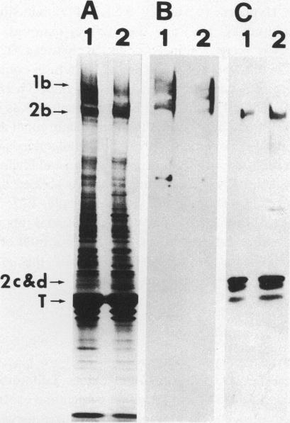
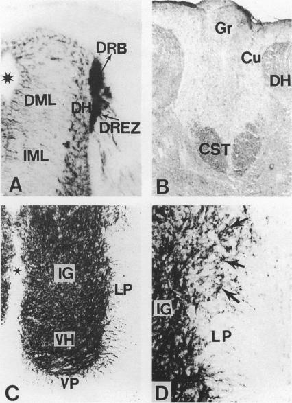
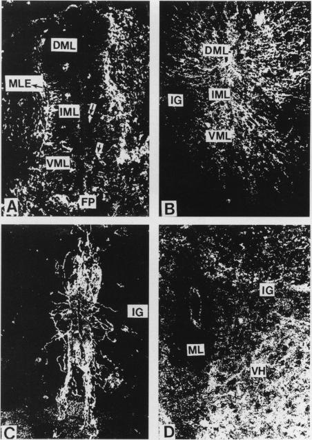
The straightforward anatomical organisation of the developing and mature rat spinal cord was used to determine and interpret the time of appearance and expression patterns of microtubule-associated proteins (MAP) 1b and 2. Immunoblots revealed the presence of MAP1b and 2 in the early embryonic rat spinal cord and confirmed the specificity of the used anti-MAP mouse monoclonal antibodies. The immunocytochemical data demonstrated a rostral-to-caudal and ventral-to-dorsal gradient in the expression of MAP1b/2 within the developing spinal cord. In the matrix layer, MAP1b was found in a distinct radial pattern distributed between the membrana limitans interna and externa between embryonal day (E)12 and E15. Immunostaining for vimentin revealed that this MAP1b pattern was morphologically and topographically different from the radial glial pattern which was present in the matrix layer between E13 and E19. The ventral-to-dorsal developmental gradient of the MAP1b staining in the spinal cord matrix layer indicates a close involvement of MAP1b either in the organisation of the microtubules in the cytoplasmatic extensions of the proliferating neuroblasts or neuroblast mitosis. MAP2 could not be detected in the developing matrix layer. In the mantle and marginal layer, MAP1b was abundantly present between E12 and postnatal day (P)0. After birth, the staining intensity for MAP1b gradually decreased in both layers towards a faint appearance at maturity. The distribution patterns suggest an involvement of MAP1b in the maturation of the motor neurons, the contralaterally and ipsilaterally projecting axons and the ascending and descending long axons of the rat spinal cord. MAP2 was present in the spinal cord grey matter between E12 and maturity, which reflects a role for MAP2 in the development as well as in the maintenance of microtubules. The present description of the expression patterns of MAP1b and 2 in the developing spinal cord suggests important roles of the two proteins in various morphogenetic events. The findings may serve as the basis for future studies on the function of MAP1b and 2 in the development of the central nervous system.
利用发育中和成熟大鼠脊髓简单的解剖结构来确定和解释微管相关蛋白(MAP)1b和2出现的时间及表达模式。免疫印迹显示早期胚胎大鼠脊髓中存在MAP1b和2,并证实了所用抗MAP小鼠单克隆抗体的特异性。免疫细胞化学数据表明,发育中的脊髓内MAP1b/2的表达呈从头至尾和从腹至背的梯度变化。在基质层,胚胎第(E)12天至E15天期间,MAP1b以独特的放射状模式分布于内界膜和外界膜之间。波形蛋白免疫染色显示,这种MAP1b模式在形态和拓扑结构上与E13天至E19天期间基质层中存在的放射状胶质细胞模式不同。脊髓基质层中MAP1b染色从腹至背的发育梯度表明,MAP1b要么密切参与增殖神经母细胞胞质延伸中的微管组织,要么参与神经母细胞有丝分裂。在发育中的基质层未检测到MAP2。在套层和边缘层,E12天至出生后第(P)0天期间大量存在MAP1b。出生后,两层中MAP1b的染色强度逐渐降低,成熟时变得很淡。分布模式表明MAP1b参与大鼠脊髓运动神经元、对侧和同侧投射轴突以及上下行长轴突的成熟过程。E12天至成熟期间,MAP2存在于脊髓灰质中,这反映了MAP2在微管发育和维持中的作用。本文对发育中脊髓中MAP1b和2表达模式的描述表明,这两种蛋白在各种形态发生事件中起重要作用。这些发现可能为未来研究MAP1b和2在中枢神经系统发育中的功能奠定基础。