Zunino S J, Singh M K, Bass J, Picker L J
Department of Pathology, University of Texas Southwestern Medical Center, Dallas 75235-9072, USA.
Am J Pathol. 1996 Aug;149(2):653-63.
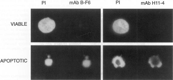
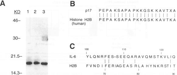
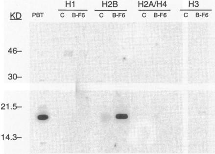
By coupling intracellular staining with terminal deoxynucleotidyl transferase (TdT)-mediated labeling of internucleosomal DNA strand breaks in a flow cytometric assay, we observed a strong correlation between apoptosis-associated DNA strand breaks and immunoreactivity with the monoclonal antibody (MAb) B-F6 in activated human peripheral blood T lymphocytes (PBTs). Although MAb B-F6 has been reported to be specific for the cytokine interleukin-6, Western blot analysis of activated PBT lysates revealed that the predominant protein band detected by this MAb was 17 kd (p17), distinct from the 23-kd core protein and 26- to 30-kd mature glycosylated forms of interleukin-6. Immunoaffinity isolation and amino-terminal amino acid sequence analysis of p17 revealed identity with the histone H2B, a finding confirmed by Western blot analysis of purified histones and by similar staining of activated PBTs with an unrelated anti-histone MAb. Neither histone staining nor DNA strand breakage was observed in freshly isolated PBTs; however, after T cell activation, histone immunoreactivity appeared to precede the appearance of DNA strand breaks, with both increasing to a maximal level by day 3 after activation. Two-parameter confocal immunofluorescence microscopy of histone and DNA staining confirmed a lack of histone immunoreactivity in viable cells and demonstrated co-localization of histone epitopes with abnormally clumped chromatin in apoptotic cells. These data indicate that alteration of histone epitope accessibility is a marker of early apoptosis and suggest that multiparameter flow cytometric analysis of intracellular epitopes may be a powerful tool in the elucidation of intracellular mechanisms of apoptosis.
通过在流式细胞术检测中,将细胞内染色与末端脱氧核苷酸转移酶(TdT)介导的核小体间DNA链断裂标记相结合,我们观察到在活化的人外周血T淋巴细胞(PBT)中,凋亡相关的DNA链断裂与单克隆抗体(MAb)B-F6的免疫反应性之间存在很强的相关性。尽管据报道MAb B-F6对细胞因子白细胞介素-6具有特异性,但对活化的PBT裂解物进行的蛋白质印迹分析显示,该单克隆抗体检测到的主要蛋白带为17kd(p17),不同于白细胞介素-6的23kd核心蛋白和26至30kd的成熟糖基化形式。对p17进行免疫亲和分离和氨基末端氨基酸序列分析,结果显示其与组蛋白H2B相同,这一发现通过对纯化组蛋白的蛋白质印迹分析以及用无关的抗组蛋白单克隆抗体对活化的PBT进行类似染色得到证实。在新鲜分离的PBT中未观察到组蛋白染色或DNA链断裂;然而,在T细胞活化后,组蛋白免疫反应性似乎先于DNA链断裂出现,两者在活化后第3天均增加到最高水平。组蛋白和DNA染色的双参数共聚焦免疫荧光显微镜检查证实活细胞中缺乏组蛋白免疫反应性,并显示凋亡细胞中组蛋白表位与异常聚集的染色质共定位。这些数据表明组蛋白表位可及性的改变是早期凋亡的一个标志物,并提示对细胞内表位进行多参数流式细胞术分析可能是阐明细胞内凋亡机制的有力工具。