Blocker A, Severin F F, Burkhardt J K, Bingham J B, Yu H, Olivo J C, Schroer T A, Hyman A A, Griffiths G
Cell Biology Programme, European Molecular Biology Laboratory, Heidelberg, Germany.
J Cell Biol. 1997 Apr 7;137(1):113-29. doi: 10.1083/jcb.137.1.113.
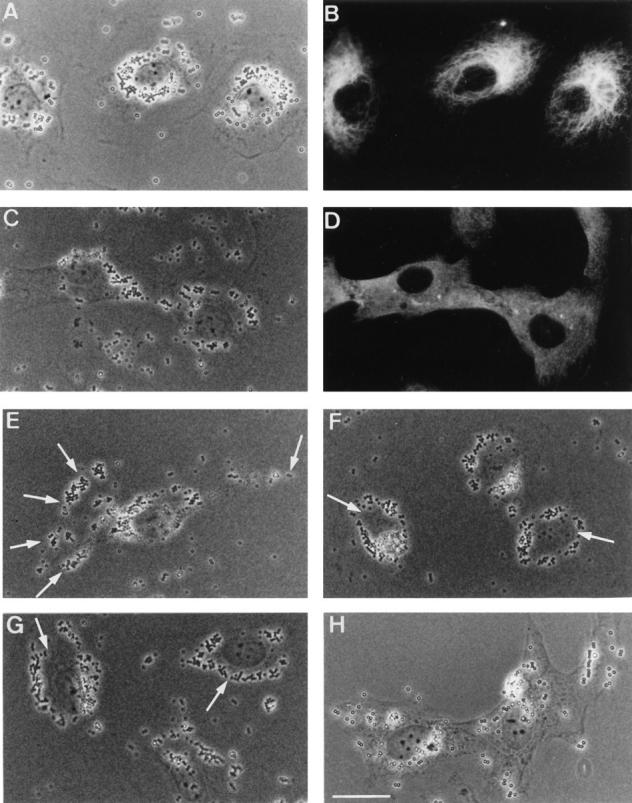
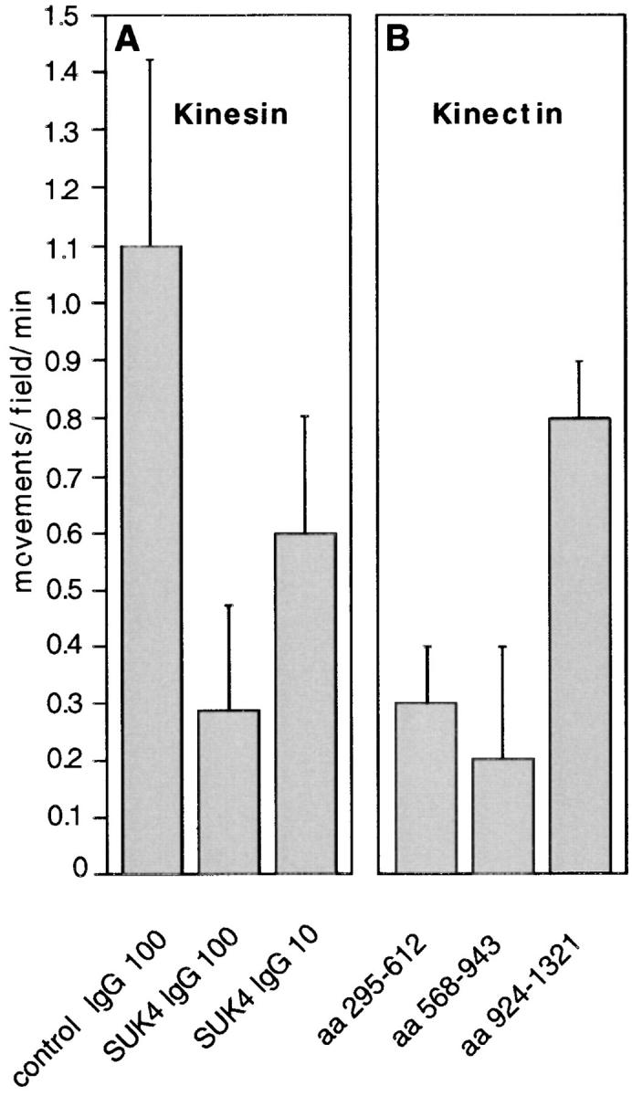
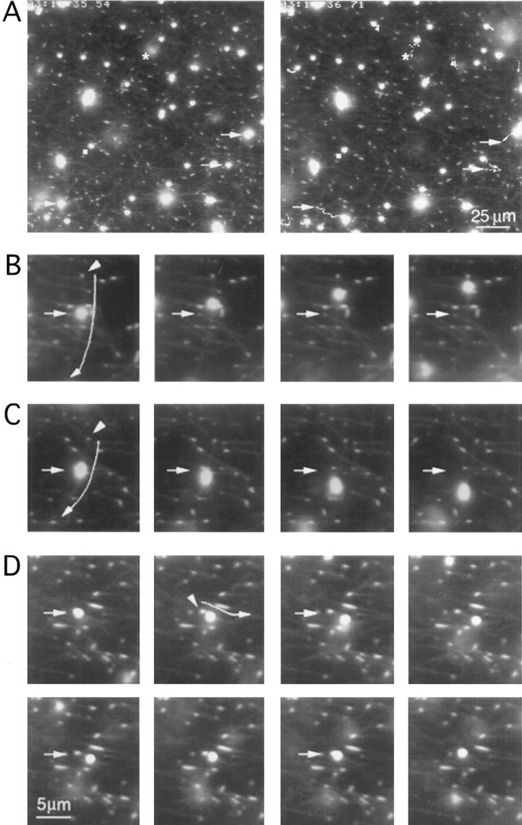
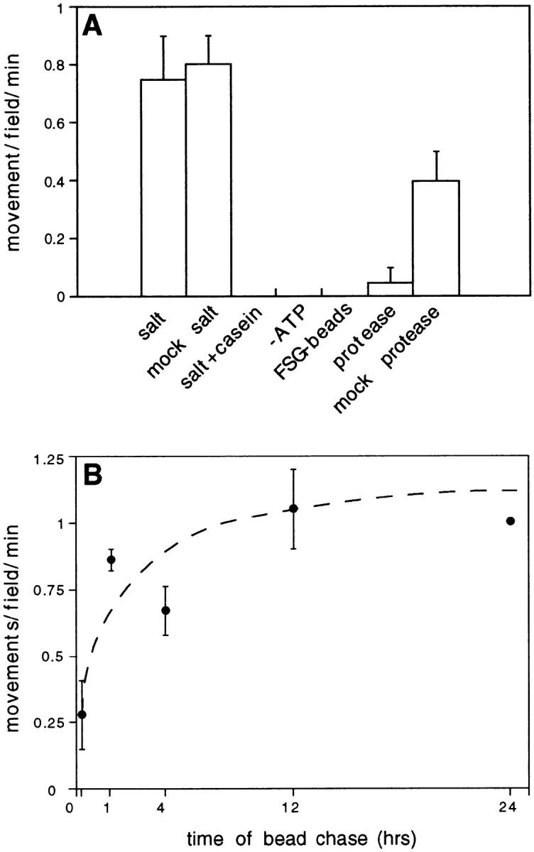
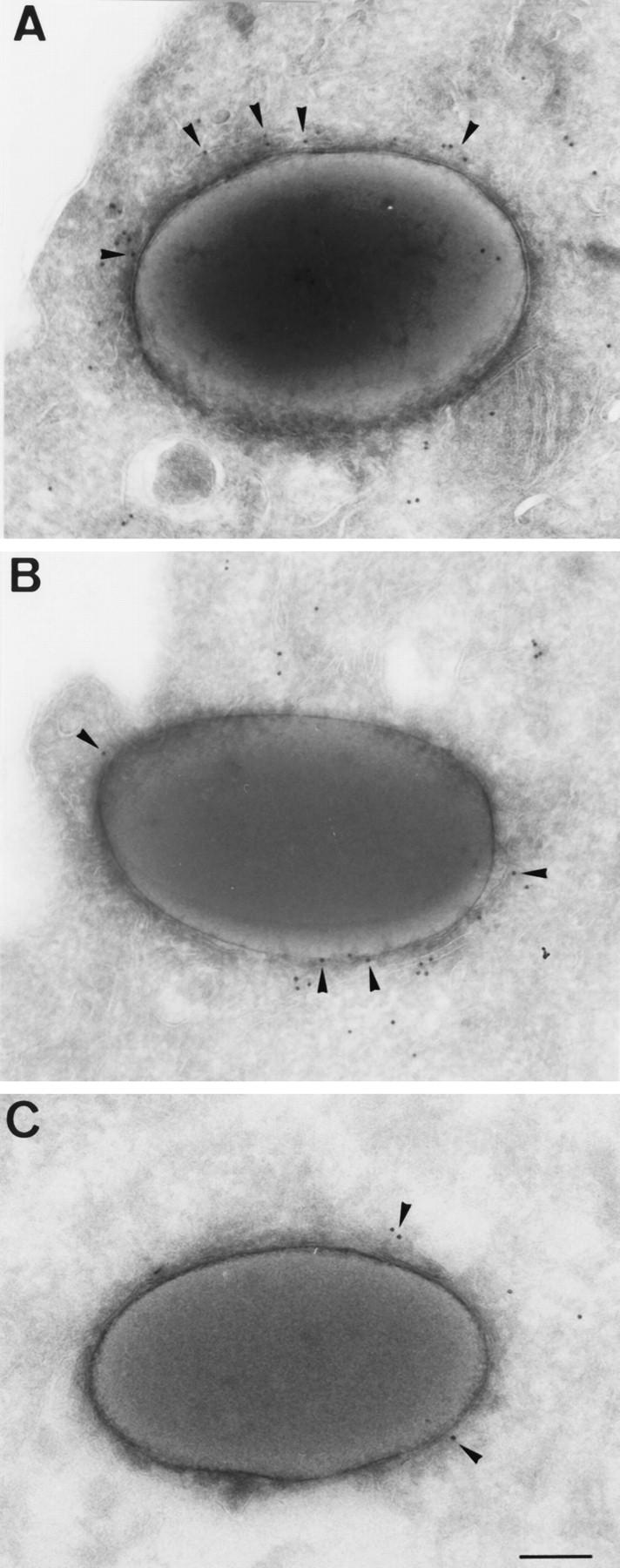
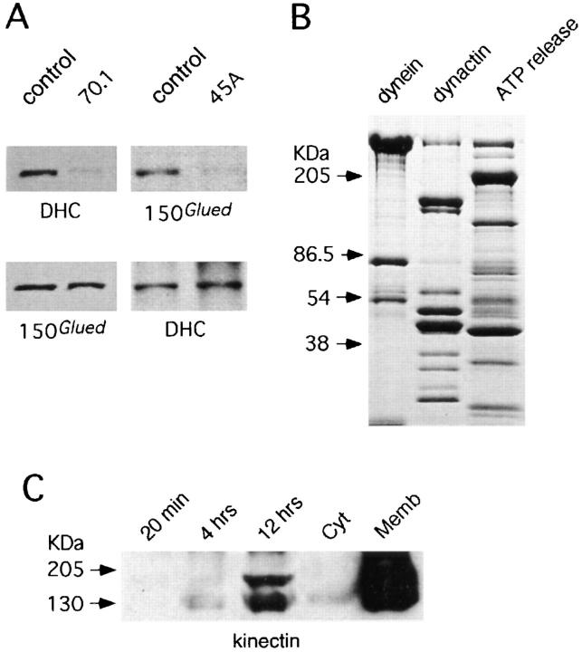
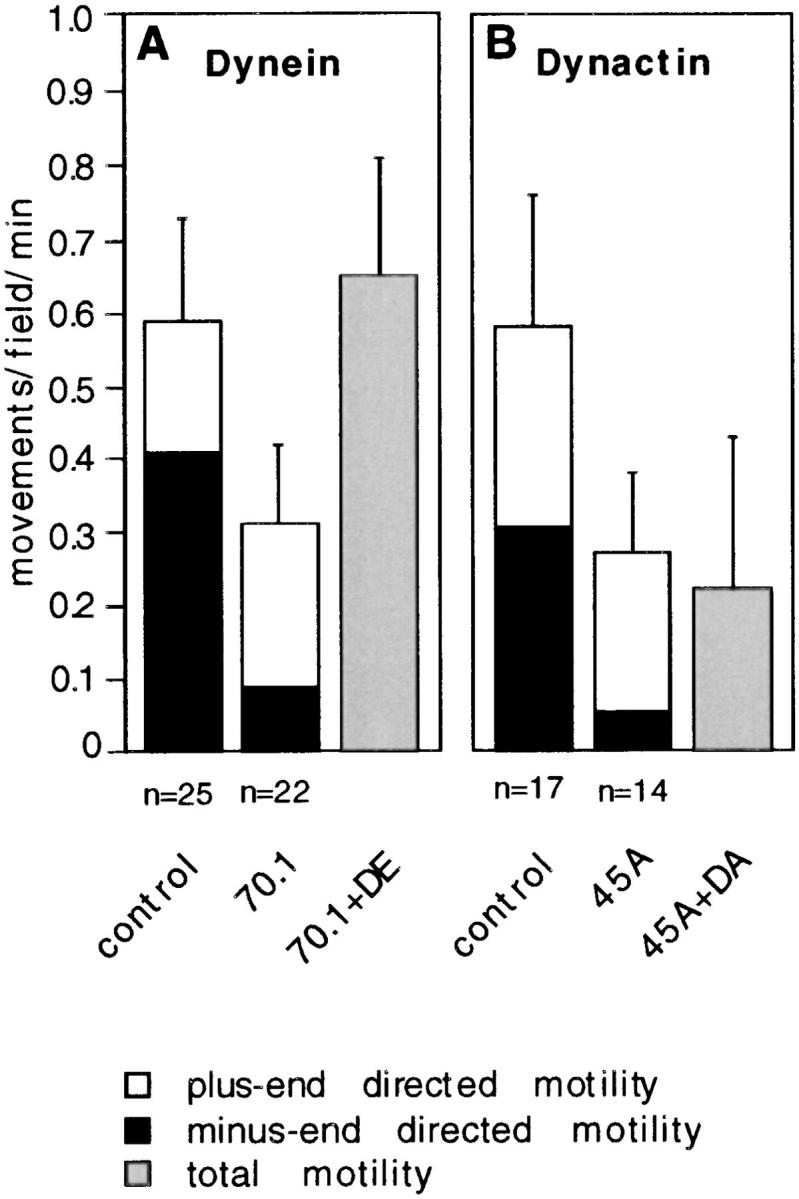
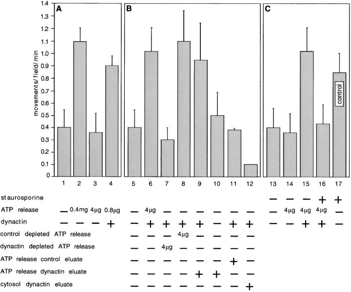
Microtubules facilitate the maturation of phagosomes by favoring their interactions with endocytic compartments. Here, we show that phagosomes move within cells along tracks of several microns centrifugally and centripetally in a pH- and microtubule-dependent manner. Phagosome movement was reconstituted in vitro and required energy, cytosol and membrane proteins of this organelle. The activity or presence of these phagosome proteins was regulated as the organelle matured, with "late" phagosomes moving threefold more frequently than "early" ones. The majority of moving phagosomes were minus-end directed; the remainder moved towards microtubule plus-ends and a small subset moved bi-directionally. Minus-end movement showed pharmacological characteristics expected for dyneins, was inhibited by immunodepletion of cytoplasmic dynein and could be restored by addition of cytoplasmic dynein. Plus-end movement displayed pharmacological properties of kinesin, was inhibited partially by immunodepletion of kinesin and fully by addition of an anti-kinesin IgG. Immunodepletion of dynactin, a dynein-activating complex, inhibited only minus-end directed motility. Evidence is provided for a dynactin-associated kinase required for dynein-mediated vesicle transport. Movement in both directions was inhibited by peptide fragments from kinectin (a putative kinesin membrane receptor), derived from the region to which a motility-blocking antibody binds. Polypeptide subunits from these microtubule-based motility factors were detected on phagosomes by immunoblotting or immunoelectron microscopy. This is the first study using a single in vitro system that describes the roles played by kinesin, kinectin, cytoplasmic dynein, and dynactin in the microtubule-mediated movement of a purified membrane organelle.
微管通过促进吞噬体与内吞区室的相互作用来促进吞噬体的成熟。在此,我们表明吞噬体在细胞内沿着数微米的轨道以离心和向心方式移动,这种移动依赖于pH值和微管。吞噬体的移动在体外得以重建,并且需要能量、该细胞器的胞质溶胶和膜蛋白。随着该细胞器的成熟,这些吞噬体蛋白的活性或存在受到调控,“晚期”吞噬体的移动频率比“早期”吞噬体高三倍。大多数移动的吞噬体是向微管负端移动的;其余的则向微管正端移动,还有一小部分双向移动。向微管负端的移动表现出动力蛋白预期的药理学特征,受到细胞质动力蛋白免疫耗竭的抑制,并且可以通过添加细胞质动力蛋白来恢复。向微管正端的移动表现出驱动蛋白的药理学特性,部分受到驱动蛋白免疫耗竭的抑制,并且通过添加抗驱动蛋白IgG完全被抑制。动力蛋白激活复合物动力肌动蛋白的免疫耗竭仅抑制向微管负端的运动性。为动力蛋白介导的囊泡运输所需的一种与动力肌动蛋白相关的激酶提供了证据。来自驱动连接蛋白(一种假定的驱动蛋白膜受体)的肽片段抑制了双向移动,该肽片段来源于与一种抑制运动性抗体结合的区域。通过免疫印迹或免疫电子显微镜在吞噬体上检测到了这些基于微管的运动因子的多肽亚基。这是第一项使用单一体外系统描述驱动蛋白、驱动连接蛋白、细胞质动力蛋白和动力肌动蛋白在纯化膜细胞器的微管介导移动中所起作用的研究。