Job C, Lagnado L
MRC Laboratory of Molecular Biology, Cambridge CB2 2QH, United Kingdom.
J Cell Biol. 1998 Dec 14;143(6):1661-72. doi: 10.1083/jcb.143.6.1661.
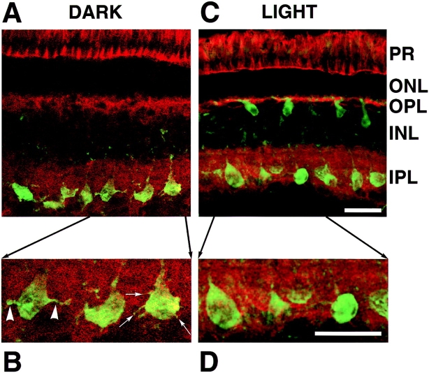
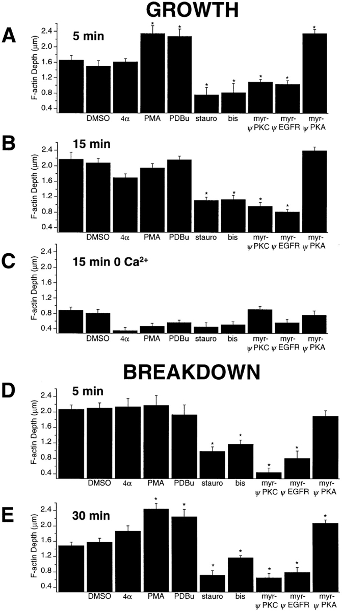
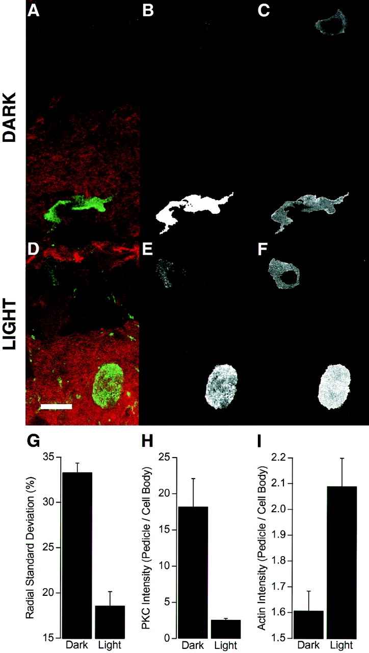
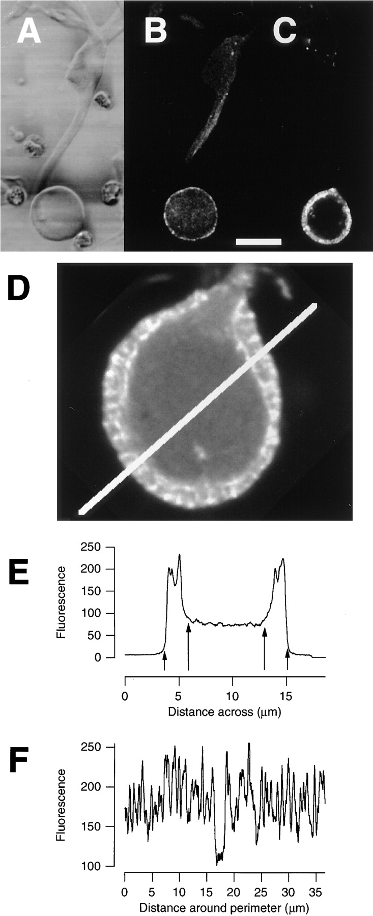
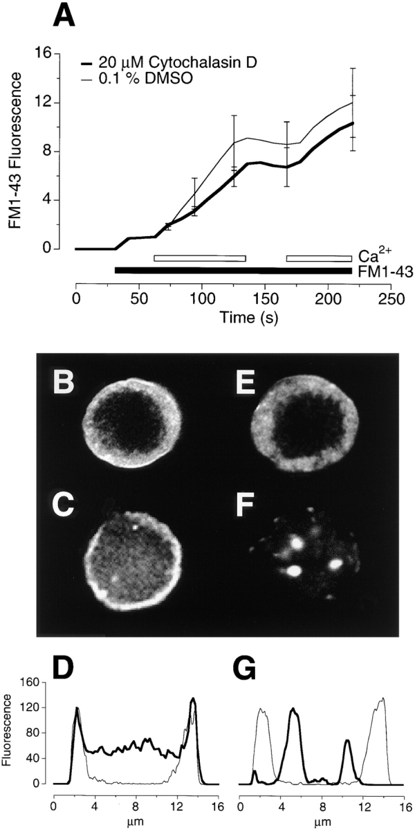
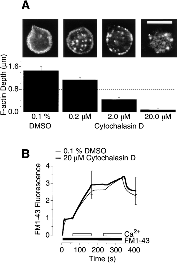
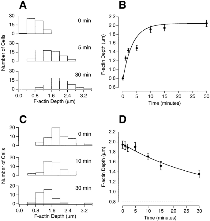
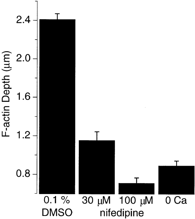
The organization of filamentous actin (F-actin) in the synaptic pedicle of depolarizing bipolar cells from the goldfish retina was studied using fluorescently labeled phalloidin. The amount of F-actin in the synaptic pedicle relative to the cell body increased from a ratio of 1.6 +/- 0.1 in the dark to 2.1 +/- 0.1 after exposure to light. Light also caused the retraction of spinules and processes elaborated by the synaptic pedicle in the dark. Isolated bipolar cells were used to characterize the factors affecting the actin cytoskeleton. When the electrical effect of light was mimicked by depolarization in 50 mM K+, the actin network in the synaptic pedicle extended up to 2.5 micrometer from the plasma membrane. Formation of F-actin occurred on the time scale of minutes and required Ca2+ influx through L-type Ca2+ channels. Phorbol esters that activate protein kinase C (PKC) accelerated growth of F-actin. Agents that inhibit PKC hindered F-actin growth in response to Ca2+ influx and accelerated F-actin breakdown on removal of Ca2+. To test whether activity-dependent changes in the organization of F-actin might regulate exocytosis or endocytosis, vesicles were labeled with the fluorescent membrane marker FM1-43. Disruption of F-actin with cytochalasin D did not affect the continuous cycle of exocytosis and endocytosis that was stimulated by maintained depolarization, nor the spatial distribution of recycled vesicles within the synaptic terminal. We suggest that the actions of Ca2+ and PKC on the organization of F-actin regulate the morphology of the synaptic pedicle under varying light conditions.
利用荧光标记的鬼笔环肽,研究了金鱼视网膜去极化双极细胞突触小蒂中丝状肌动蛋白(F-肌动蛋白)的组织情况。突触小蒂中F-肌动蛋白相对于细胞体的量,从黑暗中的1.6±0.1增加到光照后的2.1±0.1。光照还导致突触小蒂在黑暗中形成的棘状突起和突起回缩。分离的双极细胞用于表征影响肌动蛋白细胞骨架的因素。当在50 mM K⁺中通过去极化模拟光的电效应时,突触小蒂中的肌动蛋白网络从质膜延伸至2.5微米。F-肌动蛋白的形成发生在数分钟的时间尺度上,并且需要Ca²⁺通过L型Ca²⁺通道内流。激活蛋白激酶C(PKC)的佛波酯加速了F-肌动蛋白的生长。抑制PKC的试剂会阻碍F-肌动蛋白对Ca²⁺内流的生长反应,并在去除Ca²⁺后加速F-肌动蛋白的分解。为了测试F-肌动蛋白组织中依赖活性的变化是否可能调节胞吐作用或内吞作用,用荧光膜标记物FM1-43标记囊泡。用细胞松弛素D破坏F-肌动蛋白并不影响由持续去极化刺激的胞吐作用和内吞作用的连续循环,也不影响突触末端内回收囊泡的空间分布。我们认为,Ca²⁺和PKC对F-肌动蛋白组织的作用在不同光照条件下调节突触小蒂的形态。