Opella S J, Marassi F M, Gesell J J, Valente A P, Kim Y, Oblatt-Montal M, Montal M
Department of Chemistry, University of Pennsylvania, Philadelphia 19014, USA.
Nat Struct Biol. 1999 Apr;6(4):374-9. doi: 10.1038/7610.
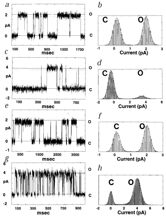
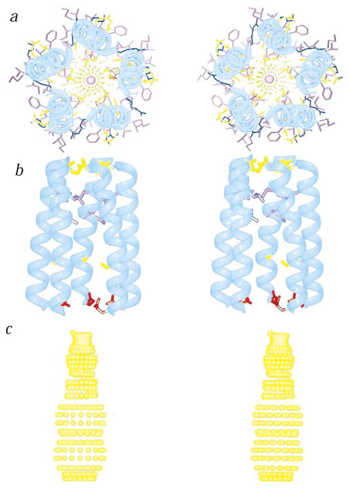
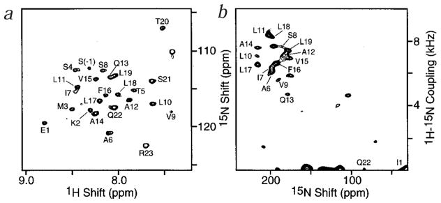
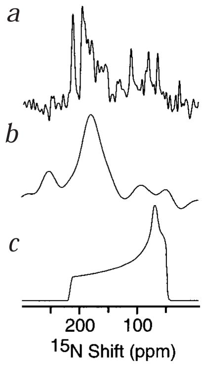
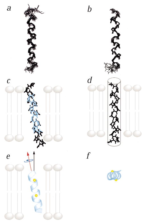
The structures of functional peptides corresponding to the predicted channel-lining M2 segments of the nicotinic acetylcholine receptor (AChR) and of a glutamate receptor of the NMDA subtype (NMDAR) were determined using solution NMR experiments on micelle samples, and solid-state NMR experiments on bilayer samples. Both M2 segments form straight transmembrane alpha-helices with no kinks. The AChR M2 peptide inserts in the lipid bilayer at an angle of 12 degrees relative to the bilayer normal, with a rotation about the helix long axis such that the polar residues face the N-terminal side of the membrane, which is assigned to be intracellular. A model built from these solid-state NMR data, and assuming a symmetric pentameric arrangement of M2 helices, results in a funnel-like architecture for the channel, with the wide opening on the N-terminal intracellular side.
利用对胶束样品进行的溶液核磁共振实验以及对双层样品进行的固态核磁共振实验,确定了与烟碱型乙酰胆碱受体(AChR)和N-甲基-D-天冬氨酸亚型谷氨酸受体(NMDAR)预测的通道内衬M2片段相对应的功能肽的结构。两个M2片段均形成无弯折的直跨膜α-螺旋。AChR M2肽以相对于双层法线12度的角度插入脂质双层,围绕螺旋长轴旋转,使得极性残基面向膜的N端侧,该侧被确定为细胞内侧。基于这些固态核磁共振数据构建的模型,并假设M2螺旋呈对称五聚体排列,结果得到一个通道的漏斗状结构,在N端细胞内侧有宽开口。